Human Reproduction, 08/07/2026
Abstract
Premature ovarian insufficiency affects 1-2% of women under 40 with limited therapeutic options beyond hormone replacement therapy, which does not restore fertility. While preclinical mouse studies demonstrate mesenchymal stem cell regenerative potential in chemotherapy-induced ovarian injury models, controlled clinical trials in human POI patients are extremely scarce, with only small uncontrolled case series reported, and the underlying cellular mechanisms remain poorly characterized.
Can human umbilical cord mesenchymal stem cells (hUC-MSCs) safely restore ovarian function in premature ovarian insufficiency (POI) patients, and what are the underlying cellular mechanisms?
Phase 1 trial demonstrated acceptable safety with significant FSH reduction and menstrual resumption; preclinical studies showed 77% follicle restoration through epithelial wound-healing mechanisms.
Methods
This study consisted of both preclinical and clinical phases.
In the preclinical phase, female ICR mice were randomly assigned to five groups (n = 10/group) and subjected to cyclophosphamide-induced ovarian injury. The animals were then treated with escalating doses of GMP-manufactured human umbilical cord mesenchymal stem cells (hUC-MSCs) derived from the Wharton’s jelly of the umbilical cord: low dose (5.33 × 10⁴ cells/mouse), moderate dose (8 × 10⁴ cells/mouse), and high dose (1.07 × 10⁵ cells/mouse). Mice were followed for 28 days, after which ovarian recovery was evaluated by histological examination with follicle counting, TUNEL assay for apoptosis, AMH immunohistochemical staining, in vitro fertilization (IVF) outcomes, and single-cell RNA sequencing (scRNA-seq) combined with CellChat analysis to investigate changes in cellular composition and intercellular communication.
The clinical phase was a single-center, open-label Phase 1 trial involving six women with premature ovarian insufficiency (POI) aged 25-40 years, defined by amenorrhea for at least four months and FSH levels > 40 mIU/mL. Each participant received a single intravenous infusion of 1.6 × 10⁸ hUC-MSCs, and was monitored for 24 weeks using a 3 + 3 dose-escalation design. Safety was assessed by recording adverse events and laboratory parameters, while preliminary efficacy was evaluated through changes in FSH, AMH, estradiol, antral follicle count (AFC), and resumption of menstruation.
Results
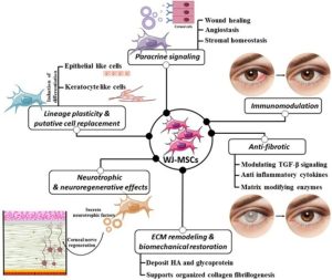
The preclinical study demonstrated that hUC-MSCs restored ovarian function in a dose-dependent manner. Compared with cyclophosphamide-injured control mice, treatment with the high dose of hUC-MSCs significantly increased the total follicle count by 34% and primordial follicle count by 77% (p < 0.001). The proportion of TUNEL-positive apoptotic cells decreased significantly from 17% to 9% (p < 0.01), while AMH expression recovered markedly (H-score: 1.0 vs. 1.9, p < 0.01). Functional recovery was also evident in in vitro fertilization (IVF) experiments, where the high-dose group achieved fertilization and blastocyst formation rates of 73.4% and 73.9%, respectively, compared with 29.6% and 29.2% in the injured control group. Single-cell RNA sequencing further revealed increased expression of wound-healing genes (Col3a1 and Col1a2) in ovarian epithelial cells, together with enhanced intercellular communication through the Angptl4-Sdc and collagen signaling pathways, promoting vascular regeneration via stromal cells.
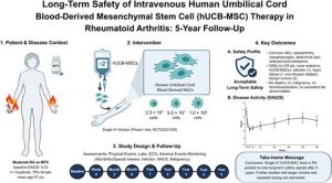
In the Phase 1 clinical trial, intravenous hUC-MSC administration demonstrated an acceptable safety profile, with no serious adverse events reported. Exploratory efficacy analysis showed a significant reduction in serum FSH levels after 24 weeks, decreasing from 82.96 ± 46.27 mIU/mL at baseline to 75.48 ± 37.88 mIU/mL (p = 0.0288). Some patients exhibited transient increases in estradiol levels exceeding 100 pg/mL, the appearance of antral follicles (mean AFC increase of 17.31 ± 21.23% at week 24), and one patient (16.7%) resumed spontaneous menstruation after six years of amenorrhea, indicating preliminary biological activity of the treatment.
Limitation
The Phase 1 clinical trial had several important limitations. The small sample size (n = 6), the absence of a control group, and the relatively short follow-up period of 24 weeks limited the ability to draw definitive conclusions regarding treatment efficacy. In addition, the substantial inter-patient variability and evaluation of only a single treatment dose indicate that larger, well-controlled studies are required to confirm these preliminary findings. Furthermore, the cyclophosphamide-induced mouse model does not fully capture the biological heterogeneity of primary premature ovarian insufficiency (POI) in humans.
Wider implications of the findings
These findings represent an important step in translating stem cell therapy for POI from promising preclinical research into early clinical application. The newly identified cellular mechanisms provide valuable insights for developing biomarkers that may help identify patients most likely to benefit from treatment. The favourable safety profile together with preliminary evidence of biological activity supports the need for larger randomized controlled trials to evaluate reproductive outcomes and fertility restoration, potentially offering a regenerative therapeutic strategy beyond the current palliative hormone replacement approach.
References
Lee, J. E., & Kim, J. H. (2026). L26/P-908 Ovarian regeneration with umbilical cord mesenchymal stem cells: preclinical evidence and phase 1 clinical trial in premature ovarian insufficiency. Human Reproduction, 41(Supplement_1), deag083-1232.
Source: Human Reproduction
Link: https://doi.org/10.1093/humrep/deag083.1232