Experimental dermatology, 06/09/2026
Introduction
Wound Healing
Wound healing consists of 4 stages: hemostasis, inflammation, proliferation, and remodeling. Hemostasis occurs immediately after injury with vasoconstriction, platelet aggregation, and the release of cytokines such as EGF, forming a fibrin blood clot that helps limit blood loss and protect against pathogens. The inflammatory stage recruits inflammatory cells and releases growth factors, cytokines, and prostaglandins, promoting signaling and angiogenesis. During the proliferation stage, angiogenesis provides oxygen, nutrients, and progenitor cells, supporting granulation tissue formation. Finally, the ECM is remodeled, blood vessels regress, and scar tissue forms.
Age, prolonged injury, and ischemia can disrupt the repair process, leading to chronic wounds. Increased levels of pro-inflammatory molecules continue to impair wound healing and increase the risk of infection and sepsis. Conventional approaches such as antibiotics, dressing changes, debridement, negative pressure, and skin grafting still have limitations such as delayed healing, scarring, and graft rejection; for diabetic foot ulcers, limb amputation may sometimes be required. Therefore, stem cell therapy has attracted attention due to its self-renewal and multipotency and its ability to promote the secretion of regenerative cytokines.
The Umbilical Cord and Umbilical Cord Blood
MSCs can be obtained from various sources such as bone marrow, umbilical cord (UC), adipose tissue, liver, and synovial tissue. Among these, the human umbilical cord is a favorable source because its collection is non-invasive, it is medical waste tissue and therefore easily obtainable, and it involves fewer ethical concerns. Umbilical cord blood (UCB) is also a rich source of stem cells and cell-secreted factors.
MSCs from UC/UCB are thought to promote wound healing through direct differentiation into cells such as endothelial cells, fibroblasts, and keratinocytes, as well as through paracrine signaling of bioactive molecules such as growth factors. hUC-MSCs have a lower degree of maturity than adult MSCs and therefore have greater self-renewal and proliferative capacities; their quantity is also less affected by the age and health status of the recipient. UCB has lower immunogenicity, with lower rates of graft rejection/graft-versus-host disease.
Recently, attention has shifted toward cell-free products from UC/UCB such as extracellular vesicles (EVs, including exosomes), conditioned medium (CM), and umbilical cord blood-derived platelet-rich plasma (UCB-PRP). These products have lower immunogenicity than cell-based products and may maintain their function during transportation and long-term storage. At the same time, the number of human clinical trials on UC/UCB products for cutaneous wound healing has been increasing. Therefore, this review focuses on synthesizing the evidence on cellular and cell-free therapies from UC/UCB, their mechanisms of action, the maturity of the clinical evidence, and the translational gaps from research to therapeutic application.
Methods
Results
Comparative Overview of UC/UCB‐Derived Cellular and Acellular Factors
Cellular Factors
Stem Cells
MSCs are multipotent cells that secrete a secretome containing bioactive molecules such as exosomes and growth factors. The most extensively studied stem cells from umbilical cord are hUC-MSCs and hUCB-MSCs; in addition, USSCs and CD34+ hematopoietic stem cells have also been studied.
Initially, it was thought that stem cells migrated to the site of injury and directly differentiated into or fused with local cells to repair the tissue. However, among 56 studies on cellular factors, only 3 preclinical studies mentioned direct differentiation. Recent evidence suggests that MSCs primarily promote skin regeneration through paracrine mechanisms, with a secretome containing growth factors, exosomes, and other bioactive molecules. The retention rate of BM-MSCs after injection into full-thickness skin wounds in diabetic mice was only 7.6% after 14 days, while USSCs only differentiated into keratinocyte-like cells in vitro, with no differentiation observed in vivo. The differentiation capacity may depend on the method of cell preparation and delivery, limiting comparisons between studies.
Acellular Factors
Cell-Free Factors
Increasing evidence suggests that paracrine mechanisms play an important, and potentially greater, role than direct differentiation in the wound-healing effects of stem cells. Therefore, cell-free factors have attracted attention because they may provide the stem cell secretome without the risks associated with cell therapy.
Extracellular Vesicles and Exosomes
Extracellular vesicles (EVs) are particles secreted by all types of living cells that function to transport proteins, lipids, and bioactive molecules. EVs can be isolated from umbilical cord blood by high-speed centrifugation and subsequently suspended in a medium such as PBS. EVs consist of 3 main subgroups: apoptotic bodies, microvesicles, and exosomes.
EVs and exosomes are the most extensively studied cell-free factors in wound healing. The most common are EVs/exosomes derived from hUC-MSCs and hUCB-MSCs; in addition, EVs from human umbilical cord blood endothelial progenitor cells (hUCB-EPCs) and mononuclear cells (hUCB-MNCs) have also been studied.
Platelet-Rich Plasma
Seven studies evaluated the potential of umbilical cord blood-derived platelet-rich plasma (UCB-PRP) in wound healing. UCB-PRP is produced by centrifuging umbilical cord blood to concentrate platelets. However, the preparation of UCB-PRP remains limited by the lack of standardized protocols, resulting in differences in composition and making it difficult to compare outcomes between studies. Although UCB-PRP contains exosomes at low concentrations, its therapeutic effects may primarily result from the large amounts of growth factors released from platelets.
Conditioned Medium
Seven studies used conditioned medium (CM) from hUC-MSCs/hUCB-MSCs for wound healing. CM contains bioactive molecules such as cytokines and exosomes, which are secreted through the paracrine mechanisms of stem cells.
Preclinical and clinical studies show that hUC-MSC CM/secretome can promote angiogenesis, cell migration, shorten wound healing time, and reduce inflammation; gels containing hUC-MSC secretome also improve chronic diabetic ulcers and trophic ulcers. However, the efficacy of cell-free factors compared with cellular factors has not been clearly established, and further clinical studies are needed to evaluate the efficacy of cell-free therapy.
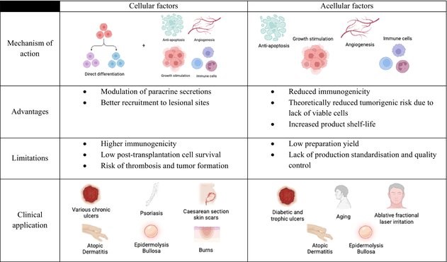
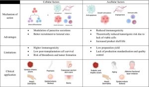
Figure 1. Comparisons between the current translational state of both cellular and acellular factors.
Preclinical Evidence
Methods of Administration and Mechanisms of Action
In preclinical animal models, both cellular and cell-free factors are mainly administered through 3 methods: topical application, intradermal injection at the injury site, or systemic intravenous injection. In addition to the direct differentiation of cellular factors, the mechanisms commonly reported to promote wound healing include immunomodulation, growth stimulation, angiogenesis, and anti-apoptosis. These mechanisms can be considered simultaneously because cell-free factors contain substances directly secreted by cells.
Immunomodulation
Cell-free factors from UC/UCB help control inflammation by reducing IL-1β, IL-6, TNF-α, and NF-κB activation, while increasing IL-4, IL-13, TGF-β, and IL-10. They promote the polarization of macrophages from M1 → M2, neutrophils from N0 → N2, and increase Treg activity. This effect is associated with miRNAs in exosomes/EVs such as miR-181c, miR-146, and miR-21, which help regulate the TLR4/NF-κB and PTEN pathways. In addition, EVs from UCB-MNCs can reduce IL-17 in a psoriasis model.
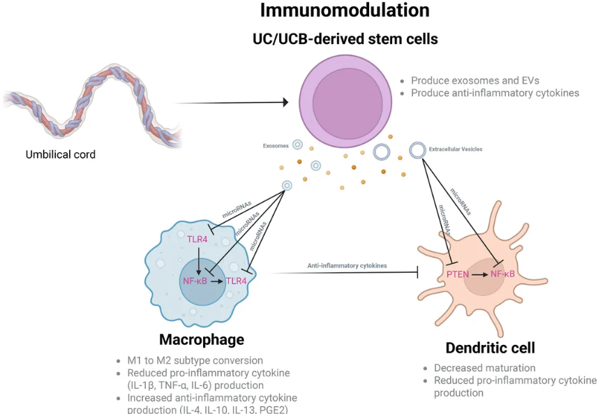
Figure 2. Summary of key cells, molecules and pathways involved in the immunomodulatory effects of UC/UCB‐derived substances to promote wound healing and skin regeneration.
Growth Stimulation
Stem cells and secretome promote wound healing by recruiting and stimulating cell proliferation. MCP-1, TGF-β, and particularly SDF-1 help attract progenitor cells and fibroblasts to the site of injury.
Secretome stimulates the proliferation of fibroblasts and keratinocytes through FGF, miR-21-3p, and the PI3K/AKT and ERK1/2 pathways. hUC-MSC exosomes also inhibit the differentiation of fibroblasts into myofibroblasts through TGF-β1/Smad2/3, contributing to limiting scar formation.
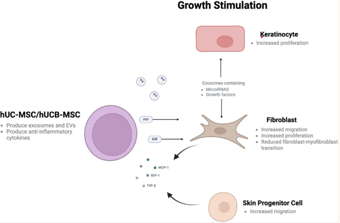
Figure 3. Summary of key cells, molecules and pathways involved in the growth‐stimulating effects of UC/UCB‐derived substances to promote wound healing and skin regeneration.
Angiogenesis
hUC-MSC exosomes promote angiogenesis by transporting Wnt4, Ang-1, Ang-2, and VEGF to the injury site, activating the Wnt/β-catenin pathway and increasing the expression of angiogenic genes. Cell-free factors also promote angiogenesis through miRNAs such as miR-135b-5p, miR-499a-3p, and miR-17-5p; among these, miR-17-5p inhibits PTEN, increases AKT/HIF-1α/VEGF activation, thereby promoting blood vessel formation.
Anti‐Apoptosis
hUC-MSC exosomes inhibit apoptosis in keratinocytes and fibroblasts through the SIRT1, ERK1/2, PI3K/AKT, p38, NF-κB, JNK, and MAPK pathways; meanwhile, miR-548ai and miR-606 inhibit ferroptosis in HaCaT cells through ACLS4. EVs from hUC-MSC apoptotic bodies also inhibit the NLRP3 inflammasome, reduce macrophage pyroptosis, and limit excessive inflammation, particularly in chronic wounds.
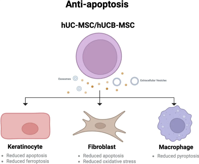
Figure 4. ummary of key cells, molecules and pathways involved in the anti‐apoptotic effects of UC/UCB‐derived substances to promote wound healing and skin regeneration.
Efficacy Across Wound Models
Traumatic Cutaneous Wounds
This is the most extensively studied model (43 studies). UC/UCB therapies generally promote full-thickness skin wound healing when administered intravenously or subcutaneously. However, the results are not entirely consistent. Doi et al. did not observe significant improvement when hUCB-MSCs were injected into 5 mm full-thickness skin wounds in nude mice. This may be related to the lower dose (1.0 × 10⁵ vs. >3.0 × 10⁵ cells) and the immunodeficient status of nude mice.
Diabetic Wounds
Twenty-four studies evaluated UC/UCB therapies for the treatment of diabetic wounds. Patients with diabetes often experience delayed wound healing due to microvascular complications, including neuropathy and reduced perfusion; there is still no definitive treatment. UC/UCB therapies showed results similar to those observed in traumatic wound models. hUC-MSC EVs promote wound healing in diabetic mice; topical application of EVs from hUC-MSC apoptotic bodies also accelerates full-thickness wound healing. In vitro, hUC-MSCs stimulate fibroblasts from patients with diabetes to increase collagen and glycosaminoglycan (GAG) secretion, contributing to improved wound healing.
Pressure Ulcers
Clinical evidence remains limited, and preclinical results are inconsistent. hUC-MSC-containing patches and UC-derived ECM hydrogels accelerate wound healing and reduce scar formation, whereas subcutaneous injection of hUC-MSCs around the wound does not significantly improve the healing rate. Similarly, hyaluronic acid hydrogel containing hUC-MSC secretome, when applied topically, improves wound healing in diabetic mice.
Burn Wounds
Burn wounds require early treatment due to the prolonged recovery process and the potential for multiple complications. All animal studies reported that cellular and cell-free factors from UC/UCB significantly accelerated burn wound healing.
Subcutaneous injection of hUC-MSC exosomes promotes the healing of second-degree burns; meanwhile, intravenous administration of hUC-MSCs in a severe full-thickness burn model improves wound healing, increases angiogenesis, and reduces inflammation. In addition, serum from burn patients can stimulate the proliferation and increase the viability of hUC-MSCs, suggesting a potential mechanism underlying the efficacy of UC/UCB-derived products in burn treatment.
Psoriasis
Three studies evaluated the therapeutic effects of UC/UCB-derived substances on psoriasis. EVs from hUCB-MNCs showed anti-inflammatory effects in vitro but did not significantly improve psoriatic manifestations in a mouse model, and therefore were proposed as an adjunctive therapy. Meanwhile, hUCB-MSCs improved psoriasis-like inflammation based on histological evaluation and flow cytometry, although no data on gross manifestations were available.
Radiation Wounds and Photodamage
Six in vitro and animal studies investigated UC/UCB therapies. Four studies showed that hUC-MSCs, hUC-MSC-derived exosomes/CM, and combined hUC-MSC/UCB-PRP therapy improved radiation-induced skin ulcers, UV-induced injury, and delayed radiation wound healing in mice. However, one study did not observe an effect of direct hUC-MSC injection, whereas hUC-MSC–SIS composite hydrogel promoted wound healing.
Epidermolysis Bullosa
Epidermolysis bullosa (EB) is a genetic disease that causes painful skin and mucosal lesions, which can lead to infection and squamous cell carcinoma. There is currently no definitive treatment; therefore, UC/UCB therapies have attracted attention.
Two studies showed that hUCB-USSCs express collagen C7, a protein that is defective in recessive dystrophic EB (RDEB); injection of hUCB-USSCs into the liver of RDEB mice improved the histology of lesions and prolonged survival. However, independent preclinical studies to confirm these findings are currently lacking.
Oral Mucositis
A study in mice with oral mucositis induced by glacial acetic acid showed that topical application of hUC-MSC EVs shortened wound healing time and reduced inflammatory mediators such as IL-6 and TNF-α.
Safety Outcomes
No adverse effects were reported in preclinical studies. hUC-MSCs were generally well tolerated: an intravenous dose of 2 × 10⁷ cells/kg in mice did not cause mortality over 23 days, whereas higher doses could cause death due to pulmonary capillary microthrombi. No acute toxicity was observed over 14 days or long-term toxicity after 12 weeks at doses up to 25 times higher than the usual clinical dose. In monkeys, a dose of 3 × 10⁷ cells/kg caused transient coma and fatigue.
hUC-MSC exosomes were well tolerated in guinea pigs, mice, and rabbits, with follow-up periods of up to 10 months. However, hUC-MSC EVs may enhance lung cancer cell growth when administered concomitantly with cancer cells; hUC-MSCs alone did not induce tumor formation but may promote the growth of pre-existing tumors.
Modifications and Delivery
Intrinsic Engineering
- Engineered EVs: Ultrasound stimulation of hUC-MSCs generates EVs with higher yields than naturally secreted EVs while maintaining their function.
- Genetic modification: hUC-MSCs expressing HOXA4 or CM carrying Wnt7a enhance wound closure; eNOS-loaded exosomes increase anti-inflammatory and angiogenic effects.
- Cell expansion: CBS increases cell proliferation and secretome production; CDM generates exosomes with greater regenerative effects. Nanofibers increase UCB-CD133+ cells by 250-fold after 10 days.
Preconditioning: Biological factors (TNF-α, TGF-β, burn serum, PRP), chemical factors (Wnt3a, CHIR99021, CoQ10, etc.), and physical conditions (hypoxia, serum/glucose deprivation, 25 mM glucose, 455 nm blue light) can enhance cell survival, proliferation, migration, and angiogenesis, and modulate the secretome/EVs.
Extrinsic Engineering:
- Delivery vehicles: Hydrogels, fibrin, chitosan, and ECM scaffolds increase the retention and stability of exosomes/secretome and maintain a favorable environment for wound healing; hydrogels are the most commonly used.
Combination therapy: Combining UC-derived products with other materials, such as collagen oligopeptides + hUC-MSC exosomes, may enhance anti-aging effects, ECM remodeling, and reduce inflammation.
Clinical Evidence
Efficacy
- Diabetic/chronic wounds: 7 studies in the past 5 years. hUC-MSC secretome improved 41 ulcers in 32 patients; hUC-MSC/PRP gel improved foot ulcers in 30 patients. A study of 108 patients showed that subcutaneous hUC-MSC injection increased wound healing, microcirculation, and granulation tissue formation.
- Procedural wounds: A study of 90 women after cesarean section found no clear improvement in wound healing or scarring.
- Burns: hUCB-MSCs improved healing of burns involving 10–25% of body surface area but increased early infections. hUC-MSC CM improved healing after CO₂ laser treatment in 23 patients.
- Psoriasis: hUC-MSCs improved PASI in 17 patients, but the disease recurred after treatment discontinuation.
- EB: Umbilical cord blood platelet gel improved wound healing in 2/3 children; hUCB-MSCs improved wound healing in 6 patients.
- Atopic dermatitis: hUCB-MSCs improved symptoms, particularly pruritus, in 34 patients; hUCB-MSC CM improved the disease in 28 patients.
Safety
Infection is a major concern; some studies reported increased wound infections, but most did not report serious adverse events. Systemic effects such as dizziness, fever, chills, and gastrointestinal disturbances were generally mild and self-limiting. A single hUC-MSC dose of ≤ 3.0 × 10⁶ cells/kg showed no safety concerns over 6 months; 59 patients had no significant adverse effects over 1 year.
However, safety evidence remains limited due to small sample sizes, follow-up periods of up to 3 years, and incomplete adverse event reporting. Immunogenicity may increase when administered into inflamed sites, following IFN-γ preconditioning, or with repeated administration.
Discussion and Unresolved Translational Gaps
Wound‐Specific Variation in Therapeutic Mechanisms
Differences in pathogenesis mean that different therapeutic mechanisms should be prioritized: diabetes – angiogenesis/immunomodulation; burns – immunomodulation; pressure ulcers – immunomodulation/growth stimulation/anti-apoptosis; psoriasis – immunomodulation; UVB injury – anti-apoptosis.
Differences Between Preclinical and Clinical Doses
Preclinical doses are generally higher, whereas clinical doses are limited by safety considerations. Differences in physiology and wound-healing mechanisms between animals and humans also affect outcomes; appropriate clinical doses need to be determined.
Standardization and Regulation
Differences among donors, manufacturing processes, contamination risks, and the lack of standardized characterization criteria reduce reproducibility. Large-scale production requires GMP, while the use of UC/UCB for skin regeneration remains largely at the experimental stage and lacks a unified standardized protocol.
Acellular Versus Cellular Factors
Clinical studies directly comparing the two groups are needed. Cell-free factors may be less immunogenic, but their efficacy, cost, storage, and manageability have not yet been fully established.
Comparisons With Standard of Care and Complementary Strategies
UC/UCB should be compared with standard treatments, and their potential for combination therapy should be evaluated. PRP has the advantages of being simple, safe, low-cost, and rich in multiple growth factors; it may be combined with UC/UCB to provide complementary/synergistic effects.
Limitations
One limitation of this scoping review is that a formal assessment of the risk of bias and quality of the included studies was not performed. This may limit the interpretation and comparison of the evidence, particularly regarding clinical efficacy and safety, making it difficult to draw definitive conclusions. To address this, a subsequent systematic review and meta-analysis comparing the evidence synthesized in this study may help provide more objective and clear conclusions.
Conclusion
Current evidence suggests the potential of cellular and cell-free factors derived from UC/UCB in wound healing, primarily through immunomodulation, growth stimulation, angiogenesis, and anti-apoptosis.
However, clinical evidence remains limited due to heterogeneity, small sample sizes, and short follow-up periods, which affect the assessment of efficacy and safety. UC/UCB therapies in dermatology remain at the research stage; large, controlled studies with long-term follow-up, together with standardized manufacturing and safety assessment, are needed. The relative efficacy of cellular versus cell-free therapies remains unclear, and therefore direct comparative studies are needed.
References
Sia, J., Chua, S., Morita, A., Lee, B., & Oon, H. H. (2026). Umbilical Cord-Derived Therapies for Wound Healing: A Scoping Review of Clinical and Translational Evidence. Experimental dermatology, 35(9), e70353.
Source: Experimental dermatology