International Journal of Nanomedicine, 09/09/2026
Introduction
Gastrointestinal (GI) diseases, including inflammatory bowel disease (IBD), gastric ulcers, small intestinal disorders, colorectal cancer (CRC), and gastric cancer (GC), remain major public health concerns. Pharmacological treatment is the primary approach for non-malignant GI diseases, whereas surgery is often required for malignant conditions. However, prolonged drug treatment may provide only temporary efficacy and cause adverse effects such as drug dependence, mucosal injury, gut microbiota disturbances, impaired nutrient absorption, and liver and kidney damage, driving the need for safer and more effective therapies.
Mesenchymal stem cells (MSCs) have attracted attention in regenerative medicine due to their self-renewal capacity, multipotency, and immunomodulatory properties. MSCs can be isolated from various tissues and generally meet the following criteria: (1) adherence to culture material surfaces; (2) expression of CD105, CD73, and CD90, with low or no expression of CD45, CD34, CD14/CD11b, CD79a/CD19, and HLA-DR; and (3) differentiation into osteoblasts, adipocytes, and chondrocytes in vitro.
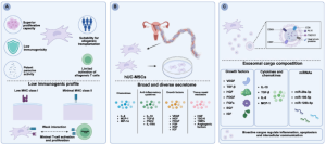
hUC-MSCs are MSCs derived from human umbilical cord tissue, offering advantages including non-invasive collection, robust ex vivo proliferation, multilineage differentiation, and low immunogenicity. Compared with BM-MSCs (bone marrow-derived MSCs) and AD-MSCs (adipose-derived MSCs), hUC-MSCs exhibit faster proliferation and stronger migratory responses toward mononuclear cells and activated lymphocytes. These properties support their potential applications in inflammatory, immune, and cancer-related diseases.
The therapeutic effects of hUC-MSCs are mainly associated with their secretome, including cytokines, growth factors, and EVs, particularly hUC-MSC-derived exosomes (hUC-MSC-exos). The secretome is influenced by the microenvironment and can be modulated through preconditioning strategies such as inflammatory cytokines, hypoxia, or 3D culture. Through paracrine mechanisms, hUC-MSCs and hUC-MSC-exos can regulate immune responses, maintain homeostasis, and promote tissue repair.
This review focuses on the biological characteristics, molecular cargo, mechanisms of action, and applications of hUC-MSCs/hUC-MSC-exos in GI diseases, while also presenting enhancement strategies and prospects for optimization and clinical translation.
Characteristics of hUC-MSCs and hUC-MSC-Exos
hUC-MSCs are multipotent stromal cells, primarily isolated from Wharton’s jelly, with advantages including high proliferative capacity, low immunogenicity, and strong paracrine activity. Compared with BM-MSCs, hUC-MSCs secrete higher levels of cytokines such as IL-6, IL-8, G-CSF, and MCP-1 and possess a richer array of active molecular components, while MMPs and ECM proteins are lower.
hUC-MSCs exhibit low MHC-I expression and minimal MHC-II/co-stimulatory molecules, making them suitable for allogeneic transplantation. hUC-MSC-exos are EVs of 30–200 nm in size, carrying markers such as CD63, CD81, CD9, ALIX, and TSG101, along with various cargos including growth factors, cytokines, and miRNAs. These cargos are involved in angiogenesis, tissue regeneration, and immune regulation, while miRNAs can regulate inflammation, apoptosis, and cancer.
Overall, the high proliferative capacity, low immunogenicity, strong paracrine activity, and cargo-delivery capability highlight the potential of hUC-MSCs/hUC-MSC-exos in tissue regeneration and immune regulation.
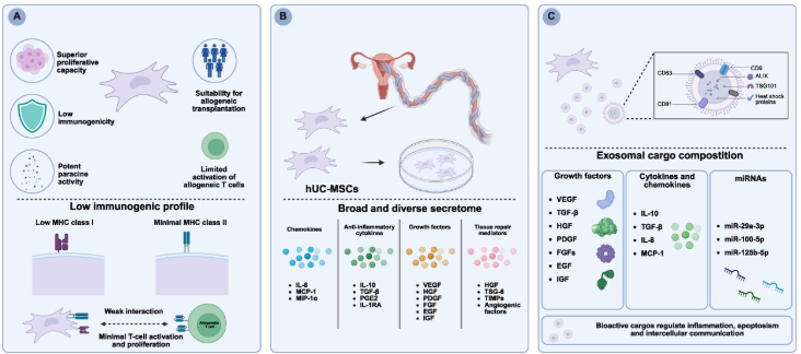
Figure 1. Characteristics of hUC-MSCs and hUC-MSC-exos. (A) Biological characteristics of hUC-MSCs; (B) Origin and secretory profile of hUC-MSCs; (C) Biological characteristics of hUC-MSC-exos.
Mechanism of hUC-MSCs and hUC-MSC-Exos for GI Diseases
Epitranscriptomic Regulation and Post-Translational Modification
The molecular cargo of hUC-MSC-exos regulates RNA epigenetics and post-translational modifications, thereby exerting therapeutic effects. In macrophages, hUC-MSC-exos increase METTL3/YTHDF1, promote m6A methylation and Slc37a2 expression, thereby enhancing M2 polarization and reducing inflammation. miR-320d from hUC-MSC-exos increases m6A and BCAT2 expression, inhibits macrophage ferroptosis, and improves IBD.
In addition to m6A, hUC-MSC-exos increase RACK1 O-GlcNAcylation, reverse EMT, reduce inflammation, and remodel the gut microbiota; meanwhile, they activate SIRT1–FXR, reduce FXR acetylation, and inhibit the NLRP3 inflammasome. In CRC, hUC-MSCs increase T-synthase and C3GnT activity, remodel O-glycans, and inhibit cancer cell proliferation and migration.
Overall, hUC-MSCs/hUC-MSC-exos regulate RNA, proteins, and inflammatory–cancer signaling, contributing to their therapeutic effects.
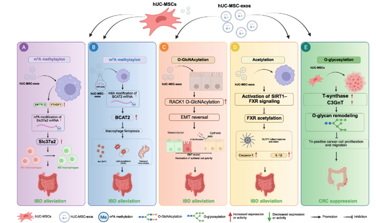
Figure 2. hUC-MSCs and their exosomes enhance tissue repair through epigenetic regulation and post-translational modifications. (A) The m6A–Slc37a2 axis in macrophages; (B) The miR-320d–BCAT2–ferroptosis axis in macrophages; (C) The O-GlcNAcylation–RACK1 axis in intestinal epithelial cells; (D) The SIRT1–FXR–NLRP3 axis in macrophages; (E) The O-glycan remodeling in CRC cells.
Modulating the Immune Response
Macrophages
hUC-MSC-exos strongly affect macrophages by regulating polarization and cell death. miR-21-5p from hUC-MSC-exos targets SPRY2, activates ERK, and promotes M2 polarization, thereby reducing colitis. miR-203a-3p inhibits Caspase-4-dependent pyroptosis; meanwhile, hUC-MSC-exos increase FXR, inhibit ferroptosis, and improve IBD. miR-378a-5p inhibits the NLRP3 inflammasome and pyroptosis, protecting against DSS-induced colitis. In CRC, miR-1827 reduces SUCNR1, inhibits tumor-promoting M2 polarization, and reduces metastasis. Overall, hUC-MSC-exos regulate context-dependent immune responses through macrophage polarization and cell death pathways.
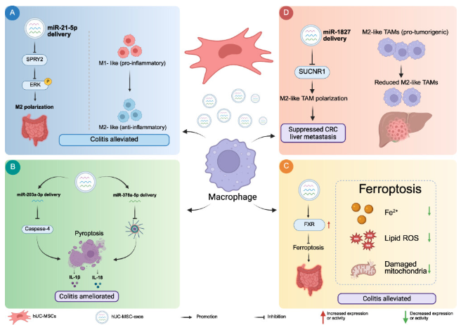
Figure 3. hUC-MSC-exos modulate macrophage responses. (A) Regulation of macrophage polarization in colitis; (B) Inhibition of macrophage pyroptosis in colitis; (C) Inhibition of macrophage ferroptosis in colitis; (D) Regulation of the tumor microenvironment in CRC.
T Cells
hUC-MSCs and hUC-MSC-exos regulate T-cell responses in IBD, mainly by balancing Treg/Tr1 and effector T cells. hUC-MSCs promote Treg differentiation through TGF-β1 while reducing Th17 responses; this mechanism is lost upon TGFB1 knockdown. In addition, hUC-MSCs promote Tr1 through an IDO-dependent mechanism while reducing Th1 and Th17. hUC-MSC-exos also shift the Treg/Th17 balance toward an anti-inflammatory state. miR-27b-3p from hUC-MSC-exos targets PIK3CA and inhibits PI3K/AKT/STAT3, restores the Th17/Treg balance, and improves DSS-induced colitis.
Other Immune Cells
In addition to macrophages and T cells, hUC-MSCs can support the generation and maturation of IL-22-producing ILC3s from CD34⁺ progenitor cells derived from umbilical cord blood. hUC-MSC-exos also protect the intestinal barrier by inhibiting mast cell activation through the IL-33/ST2 axis. Thus, hUC-MSC-derived products contribute to maintaining immune homeostasis and protecting the intestinal barrier.
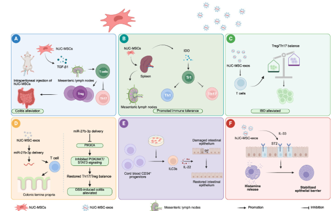
Figure 4. hUC-MSCs and their exosomes regulate immune cell function. (A) TGF-β1 – Treg/Th17 axis; (B) IDO-dependent Tr1 expansion; (C) hUC-MSC-exos shift the Treg/Th17 balance; (D) miR-27b-3p–PIK3CA–PI3K/AKT/STAT3 axis; (E) Generation of ILC3s; (F) Regulation of mast cell activation.
Regulating the Homeostasis of Gut Microbiota
hUC-MSCs and hUC-MSC-exos improve colitis through remodeling the gut microbiota and intestinal metabolism. hUC-MSCs increase SCFA-producing bacteria such as Akkermansia, Faecalibaculum, and Clostridia_UCG-014, thereby restoring immune balance. hUC-MSC-exos regulate the Firmicutes/Bacteroidetes ratio, increase beneficial bacteria, and affect the microbiota–metabolism–FXR axis, while also increasing Tregs and IgA⁺ plasma cells. Overall, they rebalance the gut microbiota, increase SCFAs, and regulate microbiota–metabolism–immune interactions, contributing to the reduction of intestinal inflammation.
Metabolic Reprogramming
hUC-MSCs and hUC-MSC-exos regulate metabolism and redox homeostasis, contributing to reduced tissue damage and tumor inhibition. In colitis, miR-129-5p/ACSL4 from hUC-MSC-exos inhibits lipid peroxidation and ferroptosis, while restoring the Xc⁻/GSH/GPX4 axis and reducing iron uptake through DMT1.
In CRC, miR-486-5p/NEK2 inhibits glycolysis and stemness, thereby reducing proliferation and metastasis. Overall, hUC-MSCs/hUC-MSC-exos reprogram metabolic–redox processes, helping reduce ferroptosis in colitis and limiting proliferative metabolism in CRC.
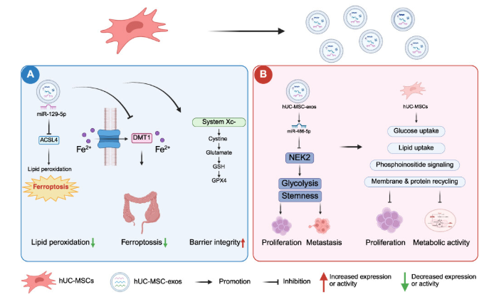
Figure 5. hUC-MSC-exos reprogram metabolic pathways. (A) Metabolic and redox regulation in colitis; (B) Metabolic regulation in CRC.
Natural hUC-MSCs and hUC-MSCs-Exos for GI Diseases
Intestinal Diseases
IBD
hUC-MSCs and hUC-MSC-exos have therapeutic potential for IBD through anti-inflammatory, tissue regenerative, immunomodulatory, and antifibrotic effects. hUC-MSCs reduce IL-1β, IL-17A, HMGB1, NLRP3, and endoplasmic reticulum stress through IRE1/XBP1. hUC-MSC-exos promote epithelial regeneration via Wnt/β-catenin, repair the mucosal barrier via TSG-6, and regulate lnc78583/miR-3202/HOXB13 and miR-302d-3p/VEGFR3/AKT. In addition, they limit fibrosis through TGF-β1/Rho/MRTF/SRF and inhibit ERK phosphorylation.
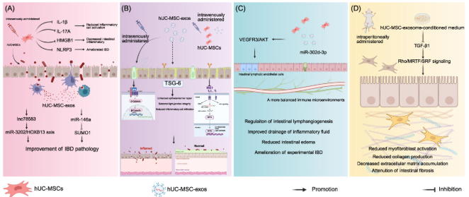
Figure 6. Roles and mechanisms of hUC-MSCs and hUC-MSC-exos in IBD. (A) Anti-inflammatory and immunomodulatory effects; (B) Intestinal epithelial regeneration and mucosal repair; (C) Lymphatic remodeling and regulation of immune homeostasis; (D) Anti-fibrotic effects.
Other Intestinal Diseases
hUC-MSCs/hUC-MSC-exos show potential in radiation-induced intestinal injury, antibiotic-induced intestinal injury, sepsis, acute pancreatitis, NEC, IBS, food allergy, and anal fistula. The main effects include reducing inflammation and apoptosis, protecting the epithelial barrier, promoting mucosal regeneration, and reducing fibrosis through mechanisms such as Nrf2/HO-1/NQO1, S100G/mTOR, miR-218-5p/CBX8, NLRP3, and HIF-1α/TGF-β/Smad.
Overall, hUC-MSCs/hUC-MSC-exos help reduce tissue injury, protect the intestinal barrier, and restore intestinal homeostasis.
Tumors
The effects of hUC-MSCs and hUC-MSC-exos on tumor progression are highly context-dependent. Some studies have shown that hUC-MSCs or hUC-MSC-exos may promote tumor growth, whereas other studies have reported tumor-inhibitory effects in certain types of cancer.
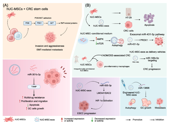
Figure 7. Roles and mechanisms of hUC-MSCs and hUC-MSC-exos in tumor therapy. (A) Pro-tumorigenic effects; (B) Anti-tumorigenic effects.
Pro-Tumorigenic Effects
In gastrointestinal cancers, hUC-MSCs and hUC-MSC-exos may promote tumor progression depending on the context. In CRC, hUC-MSCs activate PI3K/AKT, increase the expression of EMT-related proteins, and enhance invasive capacity. In gastric cancer, hUC-MSC-exos containing miR-301b-3p reduce TXNIP, promote multidrug resistance, increase cancer cell proliferation and migration, and inhibit apoptosis.
Anti-Tumorigenic Effects
hUC-MSCs and hUC-MSC-exos may inhibit tumor growth by promoting apoptosis and regulating metabolism and signaling pathways. In CRC, hUC-MSC-CM activates AMPK/inhibits mTOR, while exos carrying miR-431-5p reduce PRDX1; hUC-MSCs also reduce colitis-associated CRC and pro-inflammatory macrophages.
Engineered hUC-MSCs/exos carrying TNFSF18/CXCL9, PMO-146b, miR-655-3p, or miR-13896 have also shown inhibitory effects on CRC, ESCC, and gastric cancer. The effects depend on the cargo, cancer cells, and immune microenvironment, highlighting the need for further in vivo studies of anti-tumor immune responses. Designing defined anti-tumor cargos may help improve specificity, dose control, and safety.
Gastric Diseases
hUC-MSCs show regenerative potential in gastric mucosal diseases. In H. pylori-associated chronic atrophic gastritis (CAG), hUC-MSCs reduce inflammation, apoptosis, and mucosal fibrosis, while increasing PGI and G-17, through factors including IL-10, CD36, ATG5, STC-1, TSP-1, and 15-PGDH. hUC-MSCs also migrate to injured mucosa and promote tissue repair; submucosal injection improves CAG by inhibiting apoptosis.
In addition to CAG, hUC-MSC-CM promotes gastric ulcer healing through TRIM29/ERK/AKT, increasing cell proliferation and cell cycle progression while reducing apoptosis. Overall, hUC-MSCs exert anti-inflammatory, anti-apoptotic, antifibrotic, and mucosal regenerative effects. However, current evidence is mainly based on systemic or invasive administration routes; further studies are needed to evaluate the feasibility, efficacy, and safety of oral administration.
Strategies to Enhance the Therapeutic Potential of hUC-MSCs and hUC-MSC-Derived Exosomes
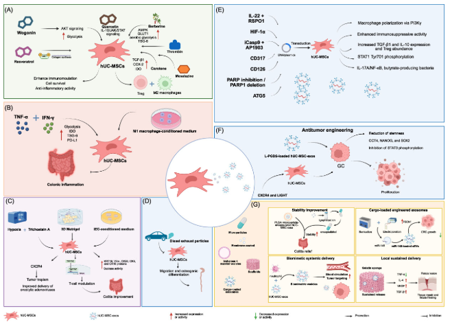
Figure 8. Strategies to enhance the therapeutic potential of hUC-MSCs and hUC-MSC-exos. (A) Small-molecule Enhanced modulation to enhance the therapeutic potential of hUC-MSCs; (B) Inflammatory priming to enhance the therapeutic potential of hUC-MSCs; (C) Culture optimization to enhance the therapeutic potential of hUC-MSCs; (D) Adverse exposure for therapeutic potential of hUC-MSCs; (E) Gene-Engineered hUC-MSCs and Exosomes for IBD; (F) Gene-Engineered hUC-MSCs and Exosomes for tumor; (G) Material Engineering and Biomimetic Delivery for hUC-MSCs and hUC-MSC-exos.
Pharmacological Preconditioning and Culture Optimization
Preconditioning with wogonin, quercetin, berberine, TNF-α/IFN-γ, and other factors can enhance the metabolism, viability, and immunomodulatory activity of hUC-MSCs, thereby improving anti-inflammatory efficacy. Hypoxia and 3D culture can also enhance targeting and immunomodulatory capacity.
Gene-Engineered hUC-MSCs and Exosomes as Targeted Therapeutic Platforms
Genetic engineering helps enhance tissue regeneration, immune regulation, and tumor targeting. Genetically modified hUC-MSCs/hUC-MSC-exos expressing IL-22/RSPO1, HIF-1α, ATG5, L-PGDS, or CXCR4/LIGHT have shown improved efficacy in IBD and tumor inhibition.
Material Engineering and Biomimetic Strategies for Optimized Delivery
Encapsulation of exosomes in PLGA, enteric delivery systems, or gelatin scaffolds, as well as the generation of cell membrane–exosome hybrids, can improve stability, oral delivery, targeting, and sustained release. Loading cargos such as miR-145 also enhances anti-CRC effects.
Overall, preconditioning, genetic modification, and biomaterial engineering enhance the activity, targeting capacity, and therapeutic efficacy of hUC-MSCs/hUC-MSC-exos.
Clinical Translation of hUC-MSCs and hUC-MSC-Exos Therapies
Initial clinical evidence suggests that hUC-MSCs and hUC-MSC-exos have therapeutic potential for refractory IBD through their immunomodulatory effects. In perianal Crohn’s disease, intrafistular injection of hUC-MSC-exos achieved complete closure in 60% of patients (12/20) and 69.7% of fistulas; hUC-MSC injection also improved disease activity and quality of life, with a 70% non-recurrence rate at 52 weeks.
In luminal IBD, intravenous hUC-MSCs demonstrated safety and the potential to improve disease outcomes. In patients with UC, clinical response and remission rates were 80.8% and 46.2%, respectively, after 2 months, and 75.0% and 37.5% after 6 months. In Crohn’s disease, combined endoscopic submucosal and intravenous administration achieved endoscopic response in 47% and mucosal healing in 17.65% of patients, while also improving intestinal barrier integrity and reducing TNF, IL-17, and TLR signaling.
Overall, initial studies indicate the feasibility, safety, and therapeutic potential of hUC-MSCs/hUC-MSC-exos in IBD, but larger randomized controlled trials are needed for confirmation.
Conclusions and Future Perspective
hUC-MSCs and hUC-MSC-exos have great potential in regenerative medicine and the treatment of gastrointestinal diseases due to their ability to provide various active molecules, including non-coding RNAs and proteins, thereby regulating immune responses, the gut microbiota, translation, and post-translational modifications. Preconditioning, genetic modification, and biomaterial engineering strategies further enhance their stability, targeting capacity, and therapeutic efficacy.
However, several challenges need to be addressed before widespread clinical application. The processes of isolation, purification, characterization, and quality control need to be standardized, while safety and therapeutic doses should be thoroughly evaluated. In cancer, because hUC-MSCs/hUC-MSC-exos may both promote and inhibit tumor growth, further studies using in vivo and organoid models are needed, particularly regarding their interactions with tumor immune cells and their potential combination with immune checkpoint inhibitor therapy.
For gastrointestinal diseases, oral administration may offer greater convenience but requires systems to protect the products from gastric acid and digestive enzymes while ensuring release at the target site. Therefore, optimization of production, stability, formulation, storage, transportation, delivery systems, and dosage is necessary. Patient-derived gastrointestinal organoids may support response assessment, dose selection, and the development of personalized therapies.
Overall, standardized production, optimized delivery, and rigorous safety evaluation are key factors for harnessing the therapeutic potential of hUC-MSCs and hUC-MSC-exos in gastrointestinal diseases.
References
Mai, Z., Yue, N., Zhao, H., Wang, L., & Li, D. (2026). Application of Human Umbilical Cord Mesenchymal Stem Cells and Their Derived Exosomes in Gastrointestinal Diseases. International Journal of Nanomedicine, 21.
Source: International Journal of Nanomedicine
Link: https://www.tandfonline.com/doi/full/10.2147/IJN.S630042