Stem Cell Reviews and Reports, 16/07/2026
Introduction
Non-alcoholic fatty liver disease (Non-Alcoholic Fatty Liver Disease – NAFLD) was previously the primary term for chronic liver disease associated with obesity and metabolic syndrome; however, as a diagnosis of exclusion, it does not fully reflect the nature of metabolic dysfunction. In 2020, the term MAFLD was proposed with positive diagnostic criteria based on metabolic abnormalities, considering the disease as a hepatic manifestation of systemic metabolic dysfunction.
Metabolic dysfunction-associated fatty liver disease (Metabolic Dysfunction-associated Fatty Liver Disease – MAFLD) is a liver injury caused by metabolic stress, closely associated with insulin resistance (IR) and genetic factors. Histopathological features include simple steatosis, MAFLD, progressive fibrosis, and cirrhosis. This process significantly increases the risk of end-stage liver complications, including liver failure and hepatocellular carcinoma. In addition, MAFLD is independently associated with a high risk of systemic complications, particularly cardiovascular disease, due to shared pathogenic mechanisms such as chronic inflammation, insulin resistance, and dyslipidemia.
Currently, MAFLD treatment mainly relies on lifestyle modifications (diet and exercise) and control of metabolic disorders. No specific drug has yet been approved by the FDA; liver transplantation is considered a definitive treatment. However, due to limitations in donor availability, surgical complications, risk of rejection, and high cost, this approach is not suitable for all patients with end-stage liver disease (ESLD). Therefore, the development of new therapeutic strategies for MAFLD is necessary.
Mesenchymal stem cells (MSCs) are multipotent stem cells capable of self-renewal and multilineage differentiation. They can differentiate into hepatocyte-like cells and, through paracrine effects, regulate immune responses, suppress inflammation and fibrosis, and promote tissue repair and regeneration. Compared with MSCs from other sources, umbilical cord-derived mesenchymal stem cells (UC-MSCs) are more widely used due to their more primitive characteristics, stronger proliferative capacity, lower immunogenicity, and superior paracrine function compared with MSCs derived from bone marrow (BM) or adipose tissue (AT). Therefore, UC-MSCs are considered an ideal cell source for MAFLD treatment.
Novel aspects of this article include:
(1) Analysis of the multi-target mechanisms of UC-MSCs (inflammation, metabolism, fibrosis).
(2) Selective evaluation of recent preclinical and clinical studies.
(3) Emphasis on translational challenges: product standardization, quality control, and long-term safety.
Pathological Mechanism of MAFLD
The pathogenesis of MAFLD is complex and involves the interaction of multiple factors. The “two-hit” hypothesis is widely accepted:
- First hit: lipid accumulation in hepatocytes due to insulin resistance (IR).
- Second hit: oxidative stress and lipid peroxidation leading to inflammation and fibrosis.
These factors activate hepatic stellate cells (HSCs), which transform into myofibroblasts, resulting in extracellular matrix (ECM) deposition and fibrosis. Key signaling pathways include: TGF-β/Smad, PDGF, CTGF, and Wnt/β-catenin.
Disease progression: hepatic fat accumulation (steatosis) → oxidative stress and inflammatory cytokines from Kupffer cells → progression to metabolic dysfunction-associated steatohepatitis (MASH) → HSC activation → collagen production and ECM deposition → fibrosis.
This mechanism provides the basis for identifying therapeutic targets of UC-MSCs.
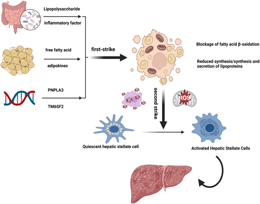
Fig 1. Pathological progression of MAFLD from steatosis to fibrosis
Characteristics and Acquisition of UC-MSCs
UC-MSCs are derived from Wharton’s jelly and perivascular tissue of the umbilical cord, with multipotent differentiation capacity, low immunogenicity, immunomodulatory properties, and paracrine effects. Compared with MSCs from bone marrow or adipose tissue, they have higher proliferative capacity and are easier to obtain. These characteristics make UC-MSCs a promising cell source for MAFLD treatment.
Mechanism of UC-MSCs in Treating MAFLD
The therapeutic mechanisms of UC-MSCs in MAFLD are not a set of isolated effects but occur in a logical sequence referred to as a “three-step cascade”:
- Step 1 – Homing & Engraftment: UC-MSCs migrate to injured liver tissue, mainly via the SDF-1/CXCR4 axis
Effective homing is a prerequisite for therapeutic success. UC-MSCs inherently express CXCR4, while SDF-1 from injured liver tissue acts as a chemoattractant, guiding the cells to the site of injury. In animal models of MAFLD, upregulation of CXCR4 using ultrasound microbubbles significantly improves the accumulation of UC-MSCs in fibrotic regions.
- Step 2 – Microenvironment Remodeling: After reaching the liver, UC-MSCs:
- Suppress inflammation (shift Kupffer macrophages from M1 → M2 phenotype, increase Treg).
UC-MSCs secrete IL-10, TSG-6 and utilize IDO1 to convert tryptophan into kynurenine → both inhibit T cell proliferation and induce differentiation of naïve T cells into Treg. At the same time, activation of the PI3K/Akt pathway promotes the transition of Kupffer cells from the pro-inflammatory M1 phenotype to the anti-inflammatory M2 phenotype, reducing TNF-α and IL-6.
- Reduce oxidative stress (via the Nrf2/HO-1 pathway)
UC-MSCs activate the Nrf2/HO-1 axis, promoting antioxidant enzymes (NQO-1, HO-1) → reducing ROS and restoring mitochondrial function. UC-MSCs can migrate to damaged intestinal mucosa and secrete TSG-6 to restore the gut barrier → reducing endotoxin (LPS) influx to the liver, thereby limiting Kupffer cell activation via TLR4/NF-κB.
- Step 3 – Restoration of Cellular Function: When the environment becomes favorable, UC-MSCs:
- Transfer mitochondria to injured hepatocytes
- Inhibit profibrotic signaling pathways
- Regulate the Bcl-2/Bax balance to reduce apoptosis
The entire process is coordinated by a paracrine network consisting of exosomes and soluble factors.
Mitochondrial Transfer—direct Metabolic Rescue
This is a significant recent advancement. In addition to paracrine mechanisms, UC-MSCs can directly transfer functional mitochondria into injured hepatocytes via two pathways: TNTs and exosomes. Studies show that mitochondria move at ~0.5 μm/s and, after 12 hours, are present in ~35% of recipient hepatocytes.
Functional effects:
- Improved hepatocyte metabolism
- Reduced fasting blood glucose (~28%) and hepatic triglycerides (~44%)
- Restored expression of fatty acid β-oxidation genes (CPT-1α, PPARα)
- Increased ATP (2.1-fold), decreased ROS (63%)
Enhancement strategies: Preconditioning UC-MSCs with healthy mitochondria increases mitochondrial transfer by ~2.5-fold, reduces the NAS score from 5.2 to 1.8—showing superior efficacy compared to conventional MSCs → Suggesting a future direction for developing mitochondria-enhanced UC-MSCs.
Anti-fibrotic and Anti-apoptotic Signaling
Even when inflammation subsides, established fibrosis does not spontaneously reverse. UC-MSCs act on hepatic stellate cells (HSCs) through at least two mechanisms:
- Anti-fibrotic: secretion of HGF and BMPs to inhibit the TGF-β/Smad pathway (the main driver of HSC activation) → reducing collagen deposition and α-SMA expression by ~50%.
- Anti-apoptotic: regulation of the Bcl-2/Bax ratio → reducing cell death; in acute liver failure models, the number of TUNEL-positive hepatocytes is reduced by more than half.
Lipid Metabolism and Insulin Sensitivity
Improving the lipid-handling capacity of hepatocytes is a key factor in functional recovery. Exosomes from UC-MSCs carry CAMKK1, which activates AMPK → inhibits SREBP-1c (the main transcription factor driving lipid synthesis) and enhances fatty acid oxidation via PPARα.
In patients with type 2 diabetes, a single infusion of UC-MSCs (1×10⁶ cells/kg) improves HOMA-IR and reduces HbA1c. However, it remains unclear whether this effect results from systemic metabolic improvement or direct hepatic action.
Non-coding RNA Networks in Exosomes—a New Layer of Regulation
In addition to protein factors, exosomes from UC-MSCs contain various regulatory RNAs capable of reprogramming the function of recipient cells. Representative molecular axes include: circ-Tulp4/miR-34a/NF-κB axis; miR-499a-5p/ETS1/GPX4 axis (ferroptosis); miR-627-5p/FTO axis. UC-MSCs not only release a mixture of biological factors but also deliver precisely regulated RNA cargos via exosomes, enabling simultaneous modulation of multiple nodes in the MAFLD pathogenic network.
Integration and Crosstalk among the Three Steps
The three steps described above do not occur in a strictly linear manner but rather interact with each other. For example, during the microenvironment remodeling phase, the released exosomes already contain miRNAs that play roles in the subsequent stage of cellular functional restoration. However, the model “homing → microenvironment remodeling → cellular functional restoration” helps explain how a single cell infusion can produce multiple therapeutic effects. At the same time, this model guides future optimization strategies, including: enhancing the homing capacity of UC-MSCs; optimizing mitochondrial transfer through cell preconditioning; engineering exosomes to enrich specific circRNAs.
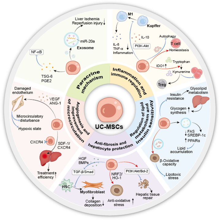
Fig 2. The “three-step cascade” mechanism of UC-MSCs in MAFLD therapy
Signaling Pathway Network Regulated by UC-MSCs
UC-MSCs treat MAFLD through a multi-target signaling network, including regulation of mitochondrial function, paracrine effects via exosomes, anti-inflammatory actions, metabolic improvement, and inhibition of fibrosis.
- Microenvironment remodeling: UC-MSCs activate Nrf2/HO-1 to reduce oxidative stress and inhibit TLR4/NF-κB through modulation of the gut–liver axis, thereby reducing inflammation.
- Restoration of cellular function: Mitochondria transferred from UC-MSCs activate AMPK and inhibit mTOR → reducing lipid synthesis and increasing fatty acid oxidation. HGF/BMPs inhibit TGF-β/Smad, thereby reducing HSC activation and ECM deposition.
- Role of exosomes: circ-Tulp4 suppresses fibrotic responses via the miR-34a/NF-κB axis; miR-499a-5p regulates ETS1/GPX4, helping to limit ferroptosis in HSCs.
In addition, UC-MSCs regulate signaling pathways related to PI3K/Akt (insulin sensitivity), Bcl-2/Bax (apoptosis), IL-6/JAK/STAT (inflammation), and Notch (cell differentiation).
Overall, the therapeutic effects of UC-MSCs in MAFLD arise from the reprogramming of the metabolic–inflammatory–fibrotic–cell fate network, rather than targeting a single pathway.
Clinical and Preclinical Research Progress
Table 1. Animal experiments and clinical studies of umbilical cord blood mesenchymal stem cells
| Study Type | Model / Population | Intervention | Main Outcomes |
| Animal Study | Mice with T2D and NAFLD model | Mitochondrial-MSCs were injected into T2D model mice via the tail vein. | Exogenous mitochondria can enhance the efficacy of MSCs in reducing blood sugar, liver transaminase, triglyceride levels and minimizing histological damage in NAFLD by mediating mitochondrial transfer. |
| Clinical Study (Retrospective) | Patients with T2DM (Type 2 Diabetes Mellitus) and MAFLD | IV infusion of UC-MSCs (50–100 × 10⁶ cells) | Significantly decreased serum levels of gamma-glutamyl transferase (GGT) and alanine aminotransferase (ALT) |
| Phase II Clinical Trial | Patients with T2DM | IV infusion of UC-MSCs (1 × 10⁶ cells/kg) | Reduced glycated hemoglobin (HbA1c) levels and improved IR |
Challenges and Future Prospects
Limitations of preclinical models and depth of mechanisms
Although preclinical studies demonstrate that UC-MSCs can reduce lipid accumulation and liver fibrosis through mechanisms such as exosome-mediated delivery of CAMKK1/miR-499a-5p and mitochondrial transfer, current animal models (HFD, MCD, CCl₄) only partially recapitulate the pathology of human MAFLD/MASH.
The mechanisms by which UC-MSCs regulate the gut–liver axis, improve systemic insulin resistance, and mediate multi-organ interactions remain unclear. In particular, although mitochondrial transfer via TNTs is a promising emerging mechanism, its efficacy and the persistence of exogenous mitochondrial function in the fibrotic liver environment in humans still require further validation.
Production processes, regulation and commercialization barriers
The widespread application of UC-MSCs still faces challenges in manufacturing standardization, quality control, and cost. Products must comply with GMP standards, with full control of the entire process from donor sourcing, cell culture, preservation to final release, to ensure consistency, purity, efficacy, and sterility.
Batch-to-batch variability must be controlled through critical quality attributes (CQAs) such as immunomodulatory capacity, exosome production, and mitochondrial function. At the same time, it is necessary to develop cell banks, serum-free media, and cell-free products (exosomes, conditioned media) to reduce costs and enhance clinical applicability.
Core obstacles in translating from animal to clinical
Although preclinical studies demonstrate the potential of UC-MSCs, clinical application still faces many obstacles. Differences in cell sources, culture conditions, isolation methods, and dosing across studies make it difficult to compare and reproduce outcomes. Currently, there is no standardized quality control framework based on the biological functions of the product.
In addition, the optimal treatment regimen has not yet been established. Studies employ various dosing levels and administration routes, but there is no consensus on dosage, route of delivery, treatment intervals, or duration. Retrospective studies mainly report improvements in biochemical parameters, while phase II trials show metabolic improvements, but their effects on liver fibrosis remain unclear.
Insufficient clinical evidence and long-term uncertainty
Current clinical data remain limited, primarily based on biochemical (ALT, GGT) and metabolic (HbA1c) markers, while lacking large-scale randomized trials assessing histological improvement in the liver; therefore, it is not yet possible to confirm whether UC-MSCs can reverse MAFLD progression. The effects across different patient populations and the predictors of treatment response have not been clearly identified. There is a need to develop biomarkers for patient selection and personalized therapy.
In addition, the long-term safety and efficacy of UC-MSCs remain unclear due to the lack of long-term follow-up data. It is necessary to assess risks such as abnormal differentiation, genomic instability, tumorigenicity, and to establish long-term monitoring systems before widespread application.
Future breakthrough directions
To overcome current challenges, future research should focus on:
(1) Using more accurate models such as organoids or humanized animal models to validate mechanisms.
(2) Standardizing the production of cell-free products such as exosomes and conditioned media.
(3) Conducting phase III clinical trials with primary endpoints of liver histological improvement, along with dose-finding studies to determine optimal regimens.
(4) Applying multi-omics technologies to identify predictive biomarkers of treatment response.
(5) Establishing long-term safety monitoring systems to evaluate efficacy and post-treatment risks.
References
Jiang, H., Wang, H., Zhu, Y. et al (2026). Human Umbilical Cord Mesenchymal Stem Cells in Metabolic Dysfunction-associated Fatty Liver Disease (MAFLD) Therapy: Mechanisms, Clinical Efficacy, and Future Perspectives. Stem Cell Rev and Rep.
Source: Stem Cell Reviews and Reports
Link: https://link.springer.com/article/10.1007/s12015-026-11181-x