International Wound Journal, 05/05/2026
Introduction
Chronic wounds, stalled in the inflammatory or proliferative phase, impose a significant global burden due to complex pathophysiological mechanisms including cellular senescence, increased pro-inflammatory cytokines, and excessive extracellular matrix–degrading enzymes (Matrix Metalloproteinases – MMPs). Conventional therapies have limited ability to improve this microenvironment; therefore, biological intervention is required.
Platelet-rich plasma (PRP) is a promising regenerative tool due to its high content of growth factors (TGF-β1, VEGF, PDGF), and has been applied in chronic ulcers, scars, and soft tissue defects. PRP can be used as monotherapy or in combination with hyaluronic acid (HA) and autologous fat grafting to enhance tissue regeneration and re-epithelialization. PRP–HA systems have demonstrated high efficacy (re-epithelialization > 96% within 30 days), even in complex wounds or those with bone exposure; the PRP–autologous fat combination leverages synergistic mechanisms between structural support and biological signaling.
However, PRP application remains controversial due to the lack of standardization in preparation protocols and treatment regimens, with many approaches requiring frequent injections that increase cost. Therefore, a standardized, minimally invasive yet effective protocol is needed.
This prospective study evaluates the efficacy and safety of a two-dose autologous PRP injection protocol in the treatment of refractory chronic non-healing wounds, aiming to determine whether this low-frequency intervention provides sufficient biological stimulation to improve wound healing over a 15-week period.
Materials and Methods
Study Design and Ethics
This was a prospective, single-arm, pre–post intervention study. All participants provided written informed consent; study records were maintained and approved by the Khon Kaen University Ethics Committee for Human Research prior to patient recruitment (Approval No.: HE641621).
Enrollment Criteria and Patient Demographics
Inclusion Criteria
Patients were included if they met the following criteria: (1) non-healing wounds refractory to standard treatment for ≥ 4 weeks; (2) contraindications to standard surgical intervention (e.g., inability to safely undergo anesthesia due to comorbidities); and (3) ankle–brachial index (ABI) ≥ 0.9.
Exclusion Criteria
Cases were excluded if they had signs of acute or systemic infection; malignancy at the wound bed; uncontrolled coagulopathy; current use of systemic corticosteroids or immunosuppressive agents; participation in another trial; or a history of allergy to blood/blood products. A total of 18 eligible patients were enrolled.
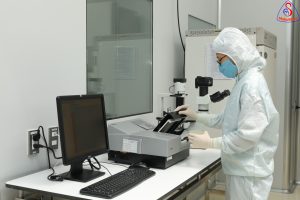
Autologous PRP Preparation Protocol
Autologous PRP was prepared from 54 mL of venous blood mixed with 6 mL of anticoagulant solution (ACD), using a two-step centrifugation protocol. The first centrifugation (1050 rpm for 10 minutes) separated the blood into three layers: platelet-poor plasma (PPP) at the top, the buffy coat containing platelets and leukocytes in the middle, and red blood cells at the bottom. The upper fraction (PPP + buffy coat) was transferred into a new sterile tube, and the red blood cell layer was discarded.
Subsequently, a second centrifugation (1400 rpm for 4 minutes) was performed to concentrate platelets into a soft pellet. Approximately two-thirds of the upper PPP layer was then removed, and the remaining portion was resuspended to obtain approximately 6 mL of final PRP. Only PRP samples achieving a post-preparation platelet concentration ≥ 4-fold higher than each patient’s baseline were used, to ensure a consistent therapeutic dose.
PRP Injection Protocol
A standardized two-dose protocol was applied: the first dose at baseline (Day 0) and the second dose after 3 weeks. After preparation, the concentrated PRP (~6 mL per dose) was drawn into a sterile syringe and used immediately, without exogenous activation (no CaCl₂ or thrombin), to allow physiological activation in situ and controlled release of growth factors upon contact with injured tissue and exposed collagen.
To preserve platelet viability and optimize growth factor retention, PRP was injected immediately after preparation using an intralesional to subcutaneous injection technique: aliquots of ~0.1 mL were injected approximately 1 cm apart along the entire wound margin; the remaining volume was injected directly into the wound bed. The second dose at week 3 was administered using the same procedure to ensure treatment consistency.
Wound Management and Measurement
Standardized Wound Care Protocol
Before each injection and measurement, standardized wound management was performed. All wounds were irrigated with normal saline solution. Where necessary, debridement was performed to remove necrotic tissue or hyperkeratotic skin to reduce the risk of secondary infection and prepare the wound bed for treatment. All wounds received regular saline dressing changes daily between follow-up visits, ensuring consistency in the baseline of wound care.
Outcome Measurements and Follow-Up Visits
Follow-Up Schedule
The PRP protocol consisted of two injections, with periodic assessments every 4 weeks over 15 weeks. The first injection was administered at baseline (day 0/week 0) after debridement and wound cleansing; the second booster injection was given at week 3 (day 21) to sustain the proliferative phase. Subsequent assessments (wound area measurement) were performed at weeks 7, 11, and 15 (end of study, including evaluation of wound healing and safety).
Wound Area Measurement
At each follow-up visit, wounds were photographed according to a standardized protocol: a digital camera was positioned at a fixed distance, with a reference ruler placed adjacent to the wound for size calibration. The images were used to calculate wound area (cm²).
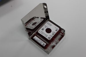
Wound area (cm²) was determined by measurements on the photographs using the reference ruler for calibration (Figure 1).

Hình 1. Size scale chart and photography of chronic wound patient.
Study Outcomes
Primary Outcome
The primary outcome was the percentage reduction in wound area (healing rate) from baseline (day 0) to week 15.
Secondary Outcomes
The documentation of systemic or local adverse events (AEs) related to the PRP injection procedure was determined. Exploration of outcomes based on associated diseases and other conditions, including radiation ulcers, venous ulcers and diabetic foot ulcers (subgroup analysis), was observed.
Statistical Analysis
Descriptive statistics were used to present sample characteristics. Wound area at day 0 and week 15 was compared using a paired t-test, with p < 0.05. The analysis was performed using SPSS.
Results
Patient Characteristics
A total of 18 patients completed the study (1 was excluded). The cohort consisted of 55.6% males and 44.4% females; 88.9% had comorbidities, predominantly diabetes mellitus. Diabetic foot ulcers were the most common (61.1%), followed by radiation-induced ulcers, venous ulcers, and the group without comorbidities (each ≤16.7%). The mean age was 57.89 ± 15.64 years; BMI was 25.45 ± 6.06. The mean duration of chronic wounds was 15.83 ± 19.05 months. The mean ABI was 1.14 ± 0.05, indicating no peripheral arterial disease.
Platelet Concentration Analysis
The effectiveness of the two-step centrifugation protocol was evaluated by measuring platelet concentrations before and after processing for both injections (Table 1). At the first injection, the baseline platelet concentration in whole blood was 250.56 ± 83.27 ×10³/µL; after concentration, PRP reached 2130.22 ± 1093.39 ×10³/µL, corresponding to an 8.5-fold increase. At the second injection (after 3 weeks), the baseline concentration was 239.61 ± 77.60 ×10³/µL; PRP after preparation reached 1682.83 ± 573.42 ×10³/µL, corresponding to a 7.0-fold increase.
Table 1. Platelet Concentration Analysis Before and After Centrifugation.
| Preparation stage | Min (103/μl) | Max (103/μl) | Mean ± SD (103/μl) | Fold increase (vs. baseline) |
| First injection | ||||
| Pre-centrifugation (baseline) | 121.00 | 384.00 | 250.56 ± 83.27 | 1.0 (Reference) |
| Post-centrifugation (PRP) | 657.00 | 4255.00 | 2130.22 ± 1093.39 | 8.5 |
| Second injection | ||||
| Pre-centrifugation (baseline) | 137.00 | 402.00 | 239.61 ± 77.60 | 1.0 (Reference) |
| Post-centrifugation (PRP) | 569.00 | 2693.00 | 1682.83 ± 573.42 | 7.0 |
The results demonstrate that the preparation protocol consistently produced therapeutic-grade PRP with high reproducibility, achieving stable elevated platelet concentrations (8.5-fold and 7.0-fold), clearly exceeding the commonly accepted therapeutic threshold (approximately 4–5-fold above baseline). Although the standard deviation of the concentrated product was relatively large, reflecting natural inter-patient variability, the mean values remained well within the therapeutic range for both doses.
Primary Outcome: Wound Area Reduction
The primary objective was to evaluate the efficacy of the two-dose PRP injection protocol in reducing the area of chronic non-healing wounds over a 15-week follow-up period.
Change in Mean Wound Area
The mean baseline wound area was 27.41 ± 70.38 cm², with a large standard deviation reflecting heterogeneity among patients. After 15 weeks of follow-up, the area decreased to 21.50 cm², corresponding to a mean reduction of 21.56%.
Paired t-test analysis showed that the reduction in wound area was statistically significant at all time points compared to baseline (p < 0.05). The most pronounced effect was observed at week 15, confirming that the two-dose PRP protocol significantly and consistently reduced wound size throughout the follow-up period.
Table 2. Changes in mean wound area over the study period (N = 18).
| Time Point | Wound area (cm2) mean ± SD | Min (cm2) | Max (cm2) | % Reduction from baseline |
| Baseline (prior to 1st PRP injection) | 27.41 ± 70.38 | 2.04 | 307.50 | – |
| Week 3 (prior to 2nd PRP injection) | 25.44 ± 70.27 | 1.50 | 304.92 | 7.19% |
| Week 7 (4 Weeks post 2nd injection) | 22.81 ± 68.52 | 0.00 | 295.36 | 16.86% |
| Week 11 (8 Weeks post 2nd injection) | 22.08 ± 68.42 | 0.00 | 294.50 | 19.52% |
| Week 15 (12 Weeks post 2nd injection) | 21.50 ± 68.97 | 0.00 | 296.40 | 21.56% |
Wound Healing Kinetics
The wound healing process was monitored periodically to evaluate the kinetics of the two-dose PRP injection protocol. After the first injection (weeks 0–3), healing progressed gradually, with a modest reduction of 7.19% prior to the second dose.
Immediately following the second injection at week 3, the rate of wound closure increased markedly, with the greatest reduction observed during the week 3–7 interval. From week 7 to week 15, healing showed a stabilizing trend, maintaining effectiveness but at a slower rate.
These kinetics suggest that the second injection plays a critical role in promoting the proliferative phase of the wound repair process.
Final Clinical Outcome and Complete Healing
At the end of the 15-week follow-up, clinical assessment showed varying degrees of treatment response. Three patients (16.67%) achieved complete re-epithelialization (100%). The majority of patients, 13 cases (72.22%), achieved partial healing with reduction in wound size and improvement in granulation tissue. Meanwhile, 2 patients (11.11%) showed poor or no response (no change or worsening).
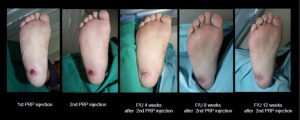
To illustrate the clinical efficacy of the two-dose PRP injection protocol, Figure 2 presents the progression of a representative good responder. A 45-year-old male patient had a diabetic right foot ulcer persisting for 3 months prior to study enrollment. Baseline images documented the ulcer at the time of the first PRP injection. By week 3 (prior to the second injection), the wound showed viable granulation tissue and margin contraction. Following the second injection, healing accelerated markedly. At 4 weeks after the second injection (week 7 of the study), the wound was nearly completely re-epithelialized with significant contraction, leaving only hyperpigmentation and scarring.

Figure 2. Resolution of a chronic diabetic foot ulcer following two-dose PRP protocol.
Subgroup Observations
Subgroup analysis showed that the two-dose PRP protocol significantly reduced wound area in most etiological groups (paired t-test, 15 weeks).
Significant effects were observed in the non-radiation ulcer group (31.34 → 25.28 cm²; p = 0.001), non-venous ulcer group (10.70 → 5.67 cm²; p = 0.003), diabetic ulcer group (11.69 → 6.77 cm²; p = 0.034), and non-diabetic group (52.12 → 44.66 cm²; p = 0.008). In contrast, radiation-induced ulcers (p = 0.387) and venous ulcers (p = 0.088) did not reach statistical significance.
Safety Profile
No serious adverse events related to blood collection or PRP injection were observed. The most common side effect was mild, transient pain at the injection site, which resolved within 24 hours. No cases of infection or systemic complications were reported, indicating a high safety profile of this autologous therapy.
Discussion
This study demonstrates that a simplified two-dose autologous PRP protocol significantly accelerates healing in chronic, difficult-to-treat wounds. The strong statistical significance confirms that the protocol generates sufficient biological signaling to shift the wound environment from a stalled inflammatory state to an active tissue remodeling phase within 15 weeks.
Efficacy and the Defined Two-Dose Protocol
The two-dose PRP protocol (Day 0 and Week 3), with high platelet concentrations (8.5×; 7.0×), demonstrated clear efficacy. Wound area decreased by 21.56% after 15 weeks, with an accelerated phase observed after the second injection (initially 7.19%).
The complete healing rate reached 16.67% in a cohort with prolonged chronic conditions, indicating that this simplified protocol remains effective, consistent, and has strong potential for broad clinical application.
Aetiology-Specific Mechanisms: Insights From Subgroup Analysis
The systematic subgroup analysis, detailed in Table 7, provides critical, clinically relevant information regarding patient selection by demonstrating a highly differential response to the PRP therapy based on the underlying wound aetiology.
Diabetic foot ulcers (DFU)
The DFU group showed the most significant improvement (p = 0.034), suggesting that PRP may overcome microvascular dysfunction and compensate for local factor deficiencies. These findings are consistent with previous studies, in which PRP monotherapy achieved 89.1% re-epithelialization within 9.7 weeks.
In this study, the longer healing time of up to 15 weeks may be attributed to the higher baseline chronicity compared to post-traumatic wounds.
Radiation Ulcers
No statistically significant improvement was observed (p = 0.387), suggesting that PRP monotherapy is limited in its ability to overcome microvascular damage and irreversible cellular loss caused by radiation injury. Previous studies indicate that more intensive combination strategies are required: PRP + HA has been shown to accelerate healing (approximately 70% re-epithelialization within 8.1 weeks), while PRP–HA scaffolds achieved up to 96.8% healing within 30 days. Therefore, for this subgroup, combining PRP with HA-based scaffolds or adipose-derived cell sources may enhance tissue regeneration.
Venous Ulcers
A trend toward improvement was observed, consistent with studies showing that PRP can reduce ulcer area within 30 days. Although the sample size was small, the pro-angiogenic effects of PRP may help alleviate hypoxia and edema associated with venous hypertension. However, long-term efficacy still depends on mechanical control (compression therapy); therefore, further studies directly comparing PRP with standard compression therapy are needed.
Strengths, Study Limitations, and Future Perspectives
Strengths: The study standardized the PRP preparation protocol with consistently high platelet enrichment (7.0–8.5-fold) and a uniform two-dose regimen. The exclusion of peripheral arterial disease (ABI > 0.9) helped reduce confounding factors.
Limitations: The absence of a randomized control group, along with a small and heterogeneous sample size (N = 18), limits generalizability, particularly across small subgroups. No adjustment for multivariate comparisons was performed, which may increase the risk of type I error. Therefore, the findings should be considered exploratory rather than confirmatory.
Conclusion
The two-dose PRP injection protocol is a safe and effective adjunctive therapy that significantly reduces wound area, particularly in diabetic ulcers. Multicenter randomized clinical trials with larger sample sizes, stratified by etiology, are needed. In addition, cost-effectiveness and quality-of-life assessments should be evaluated to support broader clinical application.
References
- Wanchutrirat, K. Jenwitheesuk, K. Jenwitheesuk, et al. (2026), Efficacy and Safety of a Defined Two-Dose Protocol of Autologous Platelet-Rich Plasma (PRP) Injection for Refractory Chronic Non-Healing Wounds: A Prospective Clinical Study, International Wound Journal 23, no. 5: e70932.
Source: International Wound Journal
Link: https://onlinelibrary.wiley.com/doi/full/10.1111/iwj.70932