Research Connections, 16/07/2026
Introduction
Airway fistulas, including tracheoesophageal fistula (TEF) and bronchopleural fistula (BPF), are serious complications associated with prolonged mechanical ventilation, thoracic surgery, malignancy, or infection. Mortality remains high due to aspiration, recurrent pneumonia, and respiratory failure. Current treatments include surgery, muscle flap reinforcement, airway stenting, or endobronchial closure techniques; most are invasive and associated with significant complication rates in frail or elderly patients.
Platelet-rich plasma (PRP) is an autologous preparation enriched with platelets in plasma. Beyond their hemostatic role, platelets are central regulators of inflammatory and immune responses. They release bioactive molecules such as PDGF, TGF-β, VEGF, and EGF, along with chemokines and cytokines, which help coordinate tissue repair and modulate inflammation.
Recent immunological evidence indicates that platelets act as “central coordinators” of the inflammatory response, with the ability to regulate leukocyte recruitment, cytokine signaling, and endothelial repair. These properties provide a mechanistic basis for the application of PRP in airway mucosal regeneration and fistula closure.
Although several case reports have described the use of PRP in airway injuries, evidence remains limited. This case series presents two detailed clinical cases and discusses the immunoregenerative basis of PRP in the treatment of airway fistulas.
Case presentation
Case 1: TEF following prolonged intubation
A 58-year-old male patient was admitted with severe community-acquired pneumonia complicated by septic shock, requiring mechanical ventilation for 21 days. The patient was intubated with an 8.0 mm endotracheal tube and had no prior history of airway disease.
During the weaning phase, the patient developed persistent coughing while eating and recurrent aspiration pneumonia. Bronchoscopy revealed a 6 mm defect in the posterior tracheal wall, consistent with a tracheoesophageal fistula (TEF). CT imaging confirmed this communication.
Due to prolonged respiratory failure, malnutrition (BMI 18 kg/m²), and poor functional reserve, the patient was not a surgical candidate. Conservative measures were ineffective.
Under bronchoscopic guidance, autologous PRP was injected circumferentially around the fistula margins (Figure 1). Two sessions were performed, 2 weeks apart. Follow-up bronchoscopy at 4 weeks demonstrated complete epithelialization and closure of the fistula (Figure 2). The patient resumed normal oral intake without recurrence.
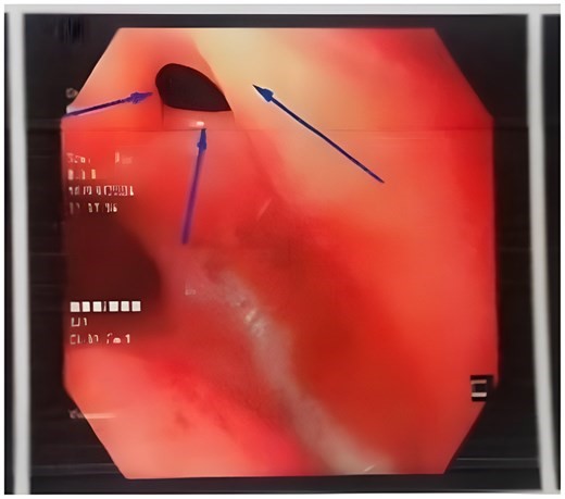
Figure 1. Bronchoscopic view of tracheoesophageal fistula before platelet-rich plasma (PRP) injection. Endoscopic image demonstrating a posterior tracheal wall defect measuring approximately 6 mm in diameter, consistent with tracheoesophageal fistula. Surrounding mucosa appears inflamed and oedematous before treatment.
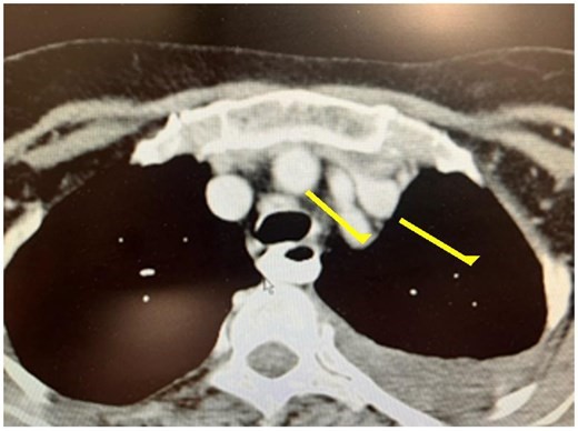
Figure 2. Axial computed tomography scan obtained four weeks after the second platelet-rich plasma injection, demonstrating complete closure of the tracheoesophageal fistula. The previous tracheoesophageal communication is no longer visible, and no evidence of persistent air leak or contrast passage is identified.
Case 2: post-lobectomy BPF
A 65-year-old female patient with severe bilateral bronchiectasis underwent right lower lobectomy due to recurrent infections. On postoperative day 9, a persistent air leak was noted. Bronchoscopy identified a 4 mm dehiscence at the bronchial stump, consistent with a bronchopleural fistula (BPF).
Due to severe chronic obstructive pulmonary disease (FEV1 = 38% predicted) and high surgical risk, the patient was not a candidate for reoperation. PRP was injected endoscopically around the bronchial defect. After two treatment sessions, the air leak resolved completely. CT imaging and bronchoscopy at 4 weeks confirmed complete healing of the lesion (Figure 3).
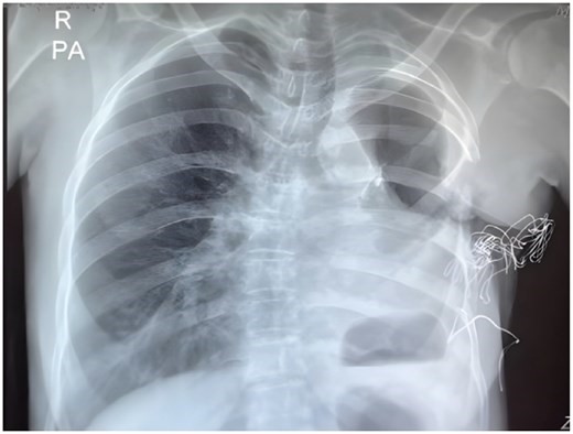
Figure 3. Post-treatment chest radiograph demonstrating resolution of the bronchopleural fistula following platelet-rich plasma therapy. No residual pneumothorax or radiographic evidence of persistent air leak is identified.
PRP preparation and treatment protocol
Autologous PRP was prepared using a double-centrifugation method. Approximately 20 mL of peripheral venous blood was collected into anticoagulant-containing tubes. The first centrifugation was performed at 1500 rpm for 10 minutes to separate plasma from red blood cells. The plasma fraction was then subjected to a second centrifugation at 3000 rpm for 10 minutes to concentrate the platelets.
The final PRP volume obtained was approximately 4–5 mL, with an estimated 4-fold increase in platelet concentration. No activation agent was used prior to injection. PRP was injected endoscopically in multiple small aliquots circumferentially around the fistula under bronchoscopic guidance. Two treatment sessions were performed, 2 weeks apart. Follow-up bronchoscopy and CT imaging were conducted 4 weeks after the second treatment session.
Discussion
This case series demonstrates the successful non-invasive closure of airway fistulas using endoscopic PRP injection in two high-risk patients who were not suitable candidates for surgery. PRP is an autologous biological product rich in platelets, containing growth factors such as PDGF, TGF-β, VEGF, and EGF, which play key roles in stimulating fibroblast proliferation, angiogenesis, and tissue regeneration. In addition to providing growth factors, platelets actively regulate immune responses and inflammatory cascades, forming the biological basis for PRP use in chronic inflammatory airway lesions.
Current treatment options for airway fistulas—including surgery, muscle flaps, stenting, and endobronchial devices—are largely invasive and often poorly tolerated in elderly or debilitated patients. PRP represents a less invasive alternative and can be repeated if necessary. However, this report does not establish definitive efficacy. Prospective clinical trials with standardized protocols are needed to evaluate safety, optimal dosing, reproducibility, and long-term effectiveness before widespread application.
Although definitive efficacy is not yet established, this study provides practical evidence supporting the feasibility of endoscopic PRP in airway fistula treatment: it presents two detailed high-risk clinical cases, reinforces the biological–immunological basis of tissue regeneration mechanisms, highlights its value in elderly/frail patients, and confirms fistula closure through imaging and endoscopy. These findings support further investigation of PRP as a minimally invasive regenerative therapy for patients who are not suitable for surgical management.
Conclusion
Endoscopic injection of autologous PRP offers a simple, safe, and effective treatment option for selected patients with tracheal or bronchial fistulas. It may serve as an alternative or adjunct to conventional surgical management, especially in high-risk or non-surgical candidates.
References
Saviz Pejhan, Iman Hesamodini, Pouyan Kamkar, Hamidreza Movahedi, Platelet-rich plasma for non-invasive closure of tracheal and bronchial fistulas: a case series, Research Connections, Volume 1, Issue 3, July 2026, vmag094.
Source: Research Connections
Link: https://academic.oup.com/rescon/advancearticle/doi/10.1093/rescon/vmag094/8736730