

Frontiers Of Oral And Maxillofacial Medicine, 28/09/2026
A preclinical study published in Frontiers of Oral and Maxillofacial Medicine in September 2026 reported that human umbilical cord-derived mesenchymal stem cells (UC-MSCs) can alleviate inflammatory lesions in the submandibular gland in a mouse model of Sjögren’s syndrome. The observed effects were particularly associated with reduced B-cell infiltration and modulation of inflammatory mediators, including HMGB1, BAFF, IL-17A, and IFN-γ.
Sjögren’s Syndrome and Exocrine Gland Lesions
Sjögren’s syndrome (SS) is a chronic autoimmune disease in which the exocrine glands, particularly the salivary and lacrimal glands, are among the most commonly affected organs. Persistent inflammation can lead to structural damage to the glands, reduced salivary secretion, and dry mouth, and may also be associated with extraglandular manifestations, including pulmonary lesions.
A key pathological feature is the accumulation of lymphocytes within glandular tissue, accompanied by increased levels of pro-inflammatory mediators. Among these, B cells and factors such as BAFF, HMGB1, IL-17A, and IFN-γ are considered to be involved in inflammatory processes and tissue injury.
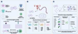
In this context, mesenchymal stem cells (MSCs) have attracted considerable interest because of their ability to modulate immune responses and control inflammation. UC-MSCs possess several characteristics that are favorable for research and the development of cell-based therapies, including robust proliferative capacity, a non-invasive tissue source, and relatively low HLA expression.
Study Design
The researchers used the NOD/Ltj mouse model, a spontaneous model commonly used to investigate pathological features resembling Sjögren’s syndrome, including lymphocytic infiltration of the salivary glands, B-cell activation, and exocrine gland dysfunction.
A total of 64 female NOD/Ltj mice were divided into groups and treated intravenously with UC-MSCs at three doses: 2.5 × 10⁵, 5 × 10⁵, and 1 × 10⁶ cells per mouse. The cells were administered on days 0, 21, and 42, and the animals were monitored for 56 days.
The researchers evaluated systemic conditions, histopathological changes in the submandibular gland, spleen, and lungs, as well as the levels of pro-inflammatory cytokines, including HMGB1, IFN-γ, IL-17A, and BAFF. The expression of several biomarkers associated with Sjögren’s syndrome, including AQP5 and KL6, was also assessed.
UC-MSCs Reduce Inflammation and Tissue Damage in the Submandibular Gland
The results showed that UC-MSC treatment markedly reduced lymphocytic infiltration in the submandibular gland. In the high-dose group receiving 1 × 10⁶ cells per mouse, the infiltration score decreased from 2.4 ± 0.5 to 1.1 ± 0.3 (p < 0.001) compared with the untreated SGL group.
At the same time, the apoptotic index in the submandibular gland decreased from 18.6 ± 3.2% to 7.3 ± 1.8% (p < 0.001) in the high-dose group. The degree of improvement showed a dose-dependent trend across the three UC-MSC doses investigated.
The observed effects were not limited to the submandibular gland. Changes were also detected in organs involved in immune responses. In the spleen and lungs, UC-MSC treatment reduced inflammatory cell infiltration, while collagen deposition in the lungs was also decreased. In the high-dose group, the lymphocytic infiltration score in the spleen decreased from 2.2 ± 0.4 to 1.0 ± 0.3, the pulmonary inflammation score decreased from 2.1 ± 0.4 to 0.9 ± 0.2, and the pulmonary fibrosis score decreased from 2.1 ± 0.4 to 1.0 ± 0.3 (all p < 0.001).
Modulation of B Cells and Pro-Inflammatory Cytokines
One of the notable findings of the study was the simultaneous modulation of B cells and inflammatory mediators.
Following treatment with high-dose UC-MSCs, serum HMGB1 levels decreased from 186.3 ± 24.5 to 98.7 ± 15.2 pg/mL; IFN-γ decreased from 156.8 ± 22.4 to 87.3 ± 14.1 pg/mL; IL-17A decreased from 89.4 ± 12.3 to 48.2 ± 8.6 pg/mL; and BAFF decreased from 1,245 ± 186 to 678 ± 95 pg/mL.
In the submandibular gland tissue, UC-MSC treatment also reduced the expression of HMGB1, BAFF, and KL6, while increasing the expression of AQP5, a protein associated with water transport and salivary secretion. In the high-dose group, the H-score for HMGB1 decreased from 210 ± 25 to 105 ± 18; BAFF decreased from 195 ± 22 to 98 ± 15; KL6 decreased from 180 ± 20 to 92 ± 14; whereas AQP5 increased from 85 ± 12 to 175 ± 20 (p < 0.001).
Two Proposed Regulatory Pathways
Based on these findings, the authors proposed that UC-MSCs may act through two interconnected regulatory pathways.
The first is the BAFF–B-cell axis. BAFF plays an important role in B-cell survival and activation. Reduced BAFF levels may therefore contribute to limiting B-cell accumulation within the submandibular gland tissue.
The second is the HMGB1–TLR4 inflammatory pathway. HMGB1 released from damaged cells can activate inflammatory signaling and subsequently promote the production of cytokines such as IFN-γ and IL-17A. The study found that UC-MSC treatment reduced HMGB1 levels together with these pro-inflammatory cytokines.
The authors suggested that these effects may involve both direct interactions between UC-MSCs and immune cells and paracrine factors secreted by UC-MSCs. However, this mechanism was inferred from in vivo findings, as the study did not include co-culture experiments to separately determine the contribution of each mechanism.
Potential and Areas for Further Research
This study provides preclinical evidence that UC-MSCs may reduce inflammatory lesions in multiple organs in the NOD/Ltj model, with effects increasing with dose within the range investigated. The dose of 1 × 10⁶ cells per mouse showed the most pronounced improvement during the 56-day observation period. However, the authors emphasized that this dose cannot yet be considered the definitive long-term optimal dose because only three dose levels were investigated and no long-term follow-up group was included.
Importantly, the study did not directly measure salivary flow. Therefore, increased AQP5 expression and improved histopathological findings are not sufficient to conclude that glandular secretory function was fully restored or that the disease was reversed. In addition, the study used only female mice, had a limited observation period of 56 days, and did not fully assess long-term efficacy or delayed adverse effects.
Conclusion
In the NOD/Ltj mouse model, intravenous administration of UC-MSCs was associated with reduced inflammatory lesions in the submandibular gland, as well as reduced inflammation in the spleen and lungs. These effects were accompanied by decreased B-cell infiltration, reduced levels of HMGB1, BAFF, IL-17A, and IFN-γ, and increased AQP5 expression in the submandibular gland.
These findings add to the preclinical evidence supporting the immunomodulatory role of UC-MSCs in autoimmune diseases and suggest the potential for further investigation of UC-MSCs in exocrine gland lesions associated with Sjögren’s syndrome. However, the current findings remain at the preclinical stage. Further mechanistic studies, direct assessment of glandular function, long-term investigations, and clinical trials are required to determine efficacy and safety in humans.
References
Zhu, Z., Fan, K., Su, C., Cheng, F., Li, X., Zeng, J., Wang, J., & Zhao, F. (2026). Human umbilical cord-derived mesenchymal stem cells alleviate lesions of the submandibular gland via B cell modulation and inflammatory cytokine suppression. Frontiers Of Oral And Maxillofacial Medicine, 0. doi:10.21037/fomm-2026-1-0003
Source: Frontiers Of Oral And Maxillofacial Medicine
Link: https://fomm.amegroups.org/article/view/125841