

Biogerontology, 25/09/2026
Introduction
MSCs are a potential cell source for tissue engineering and cell therapy due to their self-renewal, multilineage differentiation, and immunomodulatory capabilities. MSCs can be isolated from various sources, such as bone marrow, adipose tissue, dental tissue, Wharton’s jelly, and umbilical cord blood. The therapeutic potential of MSCs is primarily associated with migration to the site of injury, immunomodulation, and promotion of tissue repair through paracrine signaling.
However, MSCs can undergo cellular senescence, characterized by irreversible cell cycle arrest and the formation of SASP. Senescence reduces proliferation, migration, and osteogenic, adipogenic, and chondrogenic differentiation capacities, while pro-inflammatory factors in the SASP may sustain chronic inflammation and impair tissue regeneration.
MSCs senescence has been studied in various contexts, including donor age-related senescence, in vitro replicative senescence, and inflammation-induced senescence. The senescent state is commonly assessed using SA-β-gal, p16/p21/p53, and telomere markers. However, results across studies remain inconsistent due to differences in models, markers, and MSC tissue sources.
Objective: To systematically review the effects of cellular senescence on the functional properties of MSCs derived from adipose tissue, bone marrow, dental pulp, periodontal ligament, Wharton’s jelly, and umbilical cord blood, across different senescence contexts.
Methods
Eligibility criteria
The review was conducted according to PRISMA 2020 and the protocol was registered in PROSPERO (CRD42025638850). Research question: How does cellular senescence in MSCs from different tissues affect regenerative potential and tissue repair mechanisms?
The criteria were developed according to PECO:
- Population (P): Human MSCs derived from adipose tissue, bone marrow, dental pulp, periodontal ligament, umbilical cord blood, or Wharton’s jelly.
- Exposure (E): Cellular senescence due to donor age, repeated passaging, or inflammatory stimulation.
- Comparator (C): Non-senescent MSCs, including cells from young donors, early-passage cells, or non-stimulated cells.
- Outcomes (O): At least one characteristic related to regenerative capacity, such as proliferation, differentiation, migration, immunomodulation, or in vivo tissue repair; and the senescent state had to be confirmed by at least one marker such as SA-β-gal, p16/p21/p53, or telomere-related markers.
Only studies using primary, non-genetically modified cells were included. There were no restrictions on language or publication year.
Exclusion criteria: reviews, conference materials, case reports; studies using immortalized/genetically modified cells, lacking a non-senescent control group, or without confirmation of the senescent state.
Results
Study characteristics
The studies were published between 2006 and 2025, with a greater concentration from 2014 onward. Most studies originated from China (17; 37.8%), followed by South Korea (5; 11.1%) and the United States (4; 8.9%).
- MSC types: AD-MSCs (28%), BM-MSCs (22%), and DPSCs (22%) were the most extensively studied; UC-MSCs and PDLSCs were evaluated less frequently.
- Senescence induction models: repeated passaging was the most common (24 studies; 53%), followed by donor age (11; 24%); 7 studies (16%) evaluated both approaches. Inflammation-induced senescence was investigated in only 3 studies (4%).
- Senescence confirmation: primarily by SA-β-gal staining (91.1%), followed by p16 expression (53.3%), p21 (48.9%), and p53 (26.7%).
Functional outcomes: proliferation (86.7%) and osteogenic differentiation (73.3%) were the most commonly evaluated. In contrast, migration (6.7%), immune responses (8.9%), and in vivo tissue repair (8.9%) were assessed in fewer studies.
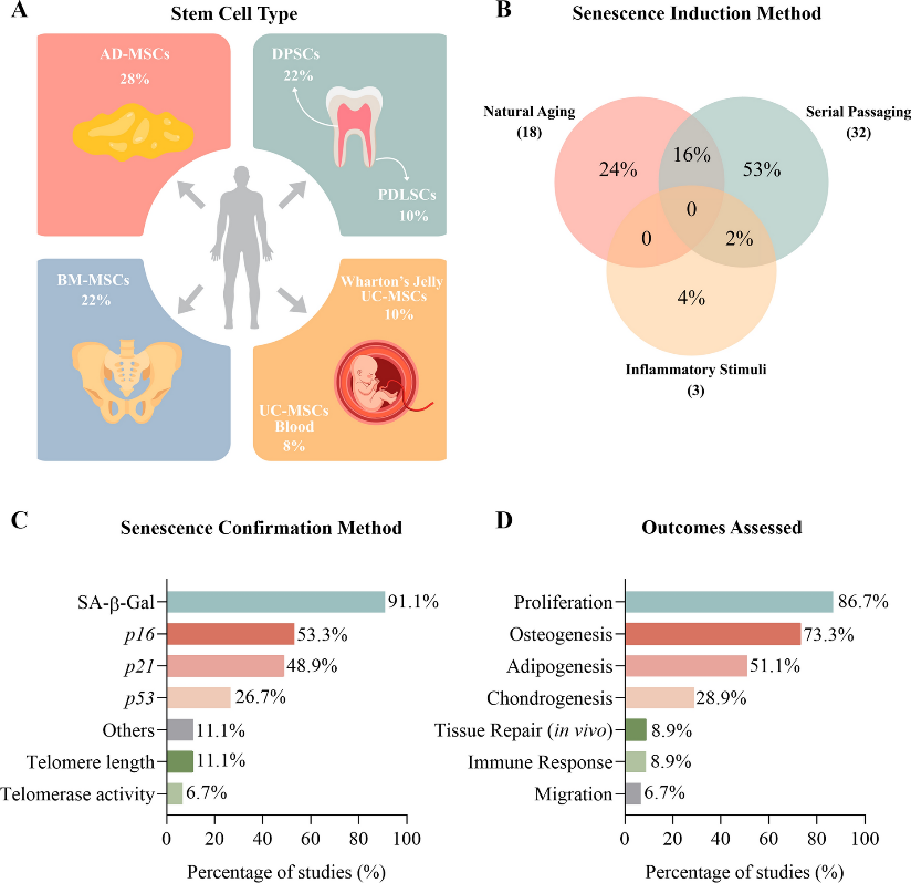
Figure 2. (A) Distribution of mesenchymal stem cell (MSC) types analyzed across studies. (B) Venn diagram of senescence induction methods. (C) Methods used to confirm cellular senescence. (D) Outcomes assessed in the included studies.
Results of individual studies
The results were synthesized according to the methods used to induce cellular senescence, including age-related senescence, repeated passaging, and inflammatory stimulation.
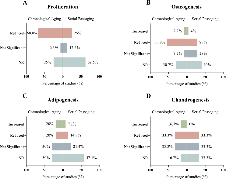
Figure 3. Comparative effects of chronological aging versus serial passaging on MSC functional capacities. Mirrored horizontal bar charts display the percentage of studies reporting increased, reduced, not significant, or not reported outcomes: A proliferation, B osteogenesis, C adipogenesis, and D chondrogenesis.
Proliferation
Inflammatory stimulation, age-related senescence, and repeated passaging are generally associated with reduced proliferative capacity of MSCs, although the results are not completely consistent across studies.
- Inflammatory stimulation: Repeated LPS stimulation reduced proliferation in DPSCs.
- Age-related senescence: Primarily reduced proliferation in AD-MSCs, BM-MSCs, and DPSCs. Some studies did not perform direct statistical comparisons.
- Repeated passaging: Had heterogeneous effects but generally showed an adverse trend in proliferation; the reduction was more evident in AD-MSCs and PDLSCs. For UC-MSCs, very few studies performed formal comparative analyses.
Studies using repeated passaging to induce senescence were included only when an independent assessment of proliferative capacity was performed, because cumulative population doublings primarily reflect replicative history rather than directly reflecting proliferative function.
Osteogenic differentiation
The effects of senescence on osteogenic differentiation were heterogeneous, depending on the method of senescence induction and the cell source.
- Inflammatory stimulation: One study on BM-MSCs and DPSCs reported a trend toward reduced osteogenic capacity in senescent cells, but this could not be confirmed due to the absence of statistical analysis.
- Age-related senescence: Most studies showed reduced osteogenic capacity in AD-MSCs, BM-MSCs, and DPSCs (7 studies; 53.8%). However, some studies reported maintained or increased differentiation capacity, depending on donor characteristics.
- Repeated passaging: The results were heterogeneous. Seven studies (28%) reported reduced osteogenic capacity in AD-MSCs, BM-MSCs, PDLSCs, and UC-MSCs; 1 study (4%) reported increased differentiation in late-passage cells, while 7 studies (28%) found no significant differences.
Overall, senescence tended to reduce osteogenic differentiation capacity, particularly in age-related senescence models, but the results remained heterogeneous across MSC sources and senescence induction methods.
Adipogenic differentiation
The effects of senescence on adipogenic differentiation were heterogeneous.
- Age-related senescence: AD-MSCs showed conflicting results, with 2 studies (20%) reporting increased and 2 studies (20%) reporting reduced adipogenic capacity. In BM-MSCs and DPSCs, most studies found no statistically significant changes or did not report changes (3 studies; 30%).
- Repeated passaging: Most studies used qualitative analyses or did not perform direct comparisons; 8 studies (57.1%) did not perform comparative statistical analyses. Among studies with comparative data, 1 study (14.3%) reported reduced and 1 study (7.1%) reported increased adipogenic differentiation capacity in senescent cells.
Overall, the effects of senescence on adipogenic differentiation remain inconsistent and may depend on the MSC source as well as the method of senescence induction.
Chondrogenic differentiation
Few studies have evaluated the chondrogenic differentiation capacity of senescent MSCs.
- Age-related senescence: Generally associated with reduced chondrogenic capacity (2 studies; 33.3%). Some studies reported maintained (2; 33.3%) or increased (1; 16.7%) differentiation capacity.
- Repeated passaging: Showed a more consistent adverse effect, reducing chondrogenic differentiation capacity in AD-MSCs, BM-MSCs, and UC-MSCs.
Overall, senescence, particularly due to repeated passaging, tended to impair chondrogenic differentiation capacity; however, the limited number of studies precludes definitive conclusions.
Migration
The migration capacity of MSCs was evaluated in very few studies. However, the available evidence indicates that both age-related senescence and repeated passaging reduced migration capacity in DPSCs, BM-MSCs, and PDLSCs (3 studies; 100%).
Immune response
Data on immunomodulatory function remain limited.
- Age-related senescence: Tended to induce a pro-inflammatory phenotype in BM-MSCs, but few studies have performed direct comparative analyses.
- Repeated passaging: Altered the expression of immune-related markers in UC-MSCs, generally toward increased levels of immunosuppressive mediators such as PGE2; however, comparative statistical analyses were not performed consistently.
Overall, the effects of senescence on immunomodulatory function remain unclear due to limited data.
In vivo tissue repair
Evidence on in vivo tissue repair capacity remains limited. Overall, inflammation-induced senescence, age-related senescence, and repeated passaging were all associated with reduced tissue regenerative capacity across multiple MSC sources. However, most studies used qualitative analyses or did not perform direct comparisons (4 studies; 80%).
Additional narrative evidence
One study on replicative senescence was not included in the summary tables because the cells were stratified based on high/low proliferative capacity rather than donor age, passage number, or inflammatory stimulation. Therefore, the results were summarized descriptively only.
- Highly proliferative cell lines maintained >80 population doublings (PDs).
- Osteogenesis: positive at 30 PDs but markedly reduced at 75 PDs.
- Adipogenesis: positive at 30 PDs and increased at 75 PDs.
All differentiation outcomes were assessed qualitatively, without statistical analysis.
Discussion
Cellular senescence is an important biological process driven by chronological aging, biological aging, and cellular stressors. In addition to its physiological roles in development and tissue homeostasis, cellular senescence is also associated with organismal aging and age-related diseases and may affect the quality of cell products used in cell therapy. However, the manifestations and responses of senescence may vary depending on the cell type.
Effects on MSC Function:
- Proliferation: This is the most consistent characteristic of senescent MSCs. Proliferative capacity is markedly reduced across all tissue sources and regardless of the senescence induction model (age-related senescence, repeated passaging, or inflammatory stress), mainly associated with activation of the p16 and p21 pathways.
- Differentiation: The effects are heterogeneous and depend on the cell source and experimental conditions. Osteogenesis may decrease, remain maintained, or increase; adipogenesis and chondrogenesis also show conflicting results. This suggests that the multilineage differentiation program may be more resilient than the cell cycle control machinery to the effects of senescence.
- Migration: Although rarely studied, the available evidence consistently indicates reduced migration capacity in DPSCs, BM-MSCs, and PDLSCs, which may affect their ability to localize to injured tissues and their regenerative efficacy.
- Immunomodulation: Senescence markedly alters the immunomodulatory phenotype, with a tendency toward increased pro-inflammatory cytokines such as TNF-α, IL-6, IL-1β, and IL-8, while anti-inflammatory factors such as IL-10, IDO-1, and TGF-β1 are regulated inconsistently. This suggests that senescence alters the secretome of MSCs toward a SASP phenotype rather than simply causing loss of immunomodulatory function.
- In vivo tissue repair: Available studies indicate that senescent MSCs have impaired tissue regenerative capacity, manifested by reduced mineralization, osteogenesis, angiogenesis, and tissue regeneration. This effect may result from impaired proliferation/differentiation combined with the adverse effects of SASP and the “bystander effect,” thereby promoting senescence in neighboring cells.
Causes of Inconsistency and Limitations of the Evidence:
Differences between studies may be related to donor age and biological characteristics, genetic background, inflammatory status, the model and extent of senescence induction, culture conditions, timing of analysis, as well as the lack of standardized criteria for defining senescence.
In addition, differences in markers and functional measurements, small sample sizes, lack of direct statistical comparisons, and data similarities among some studies limit the ability to perform quantitative synthesis. The search strategy primarily used the term “mesenchymal stem cell” rather than the ISCT-recommended term “mesenchymal stromal cell”; therefore, some studies may have been missed.
Most included studies were in vitro, with very few in vivo studies and no clinical trials. Therefore, the transferability of these findings to clinical applications remains limited. Due to the high heterogeneity, the results were primarily synthesized descriptively.
Implications for MSC Manufacturing
The consistent functional decline in senescent MSCs suggests that donor age and passage number are important quality parameters in cell manufacturing.
Changes in inflammatory phenotype and immunomodulation also highlight the need to control inflammatory stimulation during cell expansion. For MSCs intended for therapeutic use, the following should be considered:
- Screening for cellular senescence using standardized criteria;
- Assessment of the inflammatory phenotype;
- Establishment of functional quality control criteria;
- Determination of an appropriate passage number limit.
In the future, priority should be given to standardizing senescence models, directly comparing different MSC sources, increasing in vivo studies and translational models with clinically relevant outcomes, and developing strategies to prevent or reduce age-related functional decline.
Conclusion
This systematic review, based on preclinical studies, demonstrates that cellular senescence consistently affects key MSC properties related to wound healing and tissue regeneration.
- Proliferative and migratory capacities are the most markedly and consistently impaired.
- Differentiation and immunomodulatory capacities vary according to the cell source and senescence model.
- In vivo evidence indicates that senescent MSCs have reduced tissue regenerative efficacy, contributing to a better understanding of the role of senescent MSCs in organismal aging.
Although limited by methodological heterogeneity and the predominance of in vitro studies, the findings indicate that donor age, culture expansion, and inflammatory stimulation are important factors determining the functional quality of MSCs.
Therefore, standardized senescence screening and thresholds, functional quality control criteria, and passage number limits are needed to improve the safety, efficacy, and reproducibility of MSC-based regenerative therapies.
References
Watanabe, L.O., Cardoso, L.R., Silva, J.C. et al. Mesenchymal stem/stromal cell senescence in aging and regenerative medicine: a systematic review. Biogerontology 27, 167 (2026).
Source: Biogerontology
Link: https://link.springer.com/article/10.1007/s10522-026-10501-5