Journal of orthopaedic case reports, 06/2026
Introduction
Anterior cruciate ligament (ACL) reconstruction is a common procedure aimed at restoring knee stability and function following injury. Although surgical techniques have advanced, graft integration and “ligamentization” remain biologically time-dependent processes, which may delay functional recovery.
Platelet-rich plasma (PRP) has demonstrated potential and has been applied in various fields such as plastic surgery, dermatology, and osteoarthritis treatment, but it has not yet been widely used in ACL management. PRP has been investigated as a biological adjunct therapy to enhance graft healing through the delivery of concentrated growth factors. However, clinical outcomes remain inconsistent, mainly due to variations in preparation protocols, leukocyte content, and delivery techniques.
A meta-analysis has shown that PRP promotes early ligamentization and neovascularization following ACL reconstruction but does not provide significant improvement in later stages; additionally, outcomes related to postoperative pain and function remain variable. The advantages of PRP include promoting graft healing, reducing early postoperative pain and inflammation, improving tendon–bone integration, and lowering the risk of immune reactions and infection due to its autologous origin.
Leukocyte-poor PRP (LP-PRP) is believed to create a more favorable intra-articular environment by reducing pro-inflammatory cytokines while maintaining anabolic effects. Nevertheless, heterogeneity in PRP preparation and application remains a major limitation of current studies. To our knowledge, this is the first report describing a standardized LP-PRP preparation protocol combined with a systematic multi-site delivery strategy in arthroscopic ACL reconstruction.
Materials and Methods
Study population
A total of 15 patients (both male and female) who underwent arthroscopic ACL reconstruction between June 2023 and February 2024 met the eligibility criteria and were included in the study.
Study design
A total of 15 patients were included in the study.
- Inclusion criteria: patients aged ≥18 years, isolated complete ACL rupture confirmed by MRI (3 Tesla), no allergy to PRP components, underwent arthroscopic ACL reconstruction during 2023–2024, and provided informed consent.
- Exclusion criteria: active infection, partial tear, chronic ACL instability, multi-ligament knee injuries, history of autoimmune or inflammatory arthritis, coagulation disorders or current anticoagulant therapy, and systemic diseases.
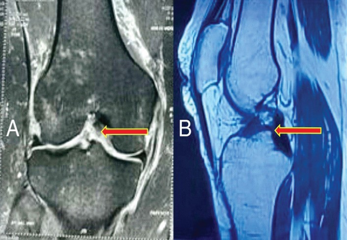
Figure 1. Pre-operative magnetic resonance imaging; (a) coronal and (b) sagittal magnetic resonance images showing disruption of the anterior cruciate ligament (arrows). There is a visible discontinuity and abnormal signal intensity of the anterior cruciate ligament consistent with tear.
Data collection
Before surgery, all patients underwent detailed medical history recording and clinical examination of the knee and lower limb. MRI was performed to confirm the diagnosis and exclude associated injuries.
After surgery, baseline functional outcomes were recorded, including IKDC score, Lysholm score, Tegner activity scale, and VAS pain score; knee range of motion (ROM) was also measured using a goniometer.
Technique
All patients underwent surgery under spinal anesthesia, with a tourniquet applied at a pressure of 350 mmHg, using the inside-out technique. Arthroscopic ACL reconstruction was performed using a quadrupled autologous hamstring graft.
An EndoButton was used for fixation at the femoral tunnel, and a bioabsorbable interference screw with a diameter 1 mm larger than the graft was used at the tibial tunnel. Fresh PRP, prepared from autologous venous blood, was used to soak the graft prior to implantation and was injected into both the tibial and femoral tunnels using a spinal needle under arthroscopic guidance at the end of the procedure.
Preparation of LP-PRP
Approximately 30 mL of autologous venous blood was collected into glass tubes containing citrate. A standardized two-step centrifugation protocol was applied based on previously published PRP methods.
The first centrifugation (soft spin) was performed at 2000 rpm for 15 minutes; the supernatant plasma layer was then aspirated under sterile conditions, avoiding the bottom red blood cell layer and the buffy coat to eliminate leukocytes. The second centrifugation (hard spin) was conducted at 3500 rpm for 10 minutes. Temperature was maintained at 20°C throughout the process to optimize platelet activation.
The upper platelet-poor plasma fraction was discarded, and the platelet pellet was resuspended to obtain LP-PRP with a high platelet concentration and minimal leukocyte contamination. Finally, calcium chloride was added to activate platelets, promoting growth factor release, initiating cell chemotaxis, and facilitating extracellular matrix deposition.
Application of LP-PRP
LP-PRP was applied using a standardized multi-site protocol based on the principle of targeted biological augmentation:
- Graft soaking: The hamstring graft was soaked in fresh LP-PRP for 10 minutes prior to implantation to enhance growth factor uptake.
- Tunnel injection: LP-PRP was injected into the femoral socket and tibial tunnel around the graft after fixation, targeting the bone–tendon interface to enhance healing.
- Intra-articular injection: The remaining PRP was injected around the intra-articular portion of the graft at the end of surgery to promote synovial and intra-articular tissue healing.
This approach ensures comprehensive exposure of the graft and surrounding tissues to the biological augmentation effects.
Rehabilitation
In the early phase (0–6 weeks), the focus was on pain control, early mobilization, and gentle ROM exercises, with weight-bearing as tolerated by the patient.
From 6–12 weeks, strengthening of the quadriceps, hamstrings, and calf muscles was emphasized through both open and closed kinetic chain exercises. Proprioceptive and functional training were also implemented to improve balance, coordination, and neuromuscular control.
After 12 weeks, sport-specific exercises and techniques were introduced to facilitate a safe return to the pre-injury level of activity.
Follow-up protocol
Primary functional outcomes (IKDC, Lysholm, Tegner scores) were assessed at 4 weeks, 8 weeks, and 6 months after surgery.
Secondary outcomes, including graft integration and healing, were evaluated through clinical examination and VAS pain scores to determine the long-term efficacy and safety of LP-PRP augmentation.
Statistical analysis
Descriptive statistical analysis was performed.
Results
A total of 15 patients were included in the study, comprising 8 females and 7 males. The mean age of the study population was 29.26 ± 8.25 years, and the mean time to return to pre-injury activity level was 5.33 ± 0.83 months.
The mean IKDC score increased from 53.86 ± 0.63 in the immediate postoperative period to 66.73 ± 1.48 at 6 months. Similarly, the Lysholm score improved from 62.06 ± 1.33 to 83.67 ± 1.83; the Tegner activity score increased from 3.40 ± 0.5 to 4.73 ± 0.7; while the VAS score decreased from 5.26 ± 0.7 to 1.60 ± 0.63 over the same period.
The mean time to achieve 90° knee flexion and full knee extension was 23.27 ± 1.09 days and 41.86 ± 1.5 days, respectively.
These results indicate improved early recovery with biological augmentation. No complications such as infection, excessive inflammation, or PRP-related adverse reactions were observed.
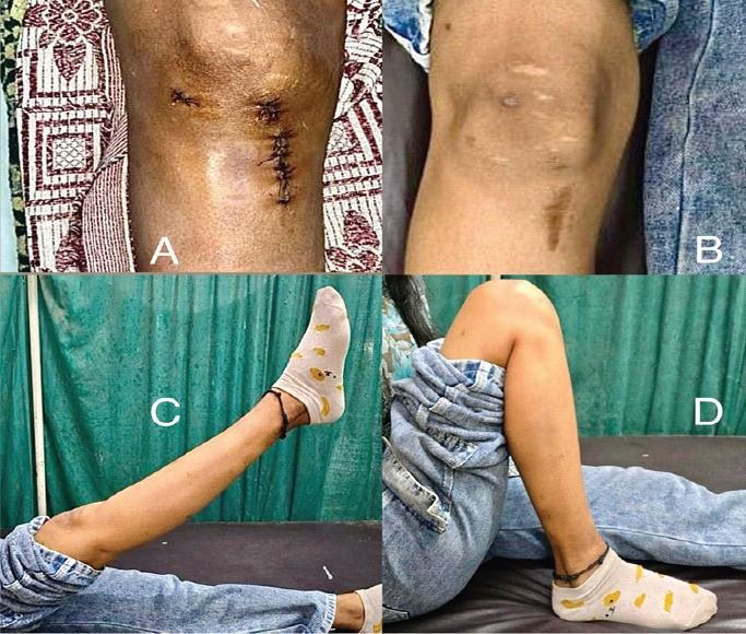
Figure 2. Clinical photos of the suture site and range of motion of the operated knee; Suture site in the (a) immediate post-operative period and (b) at 6 months follow-up. No wound-related complications were observed. Normal knee range of motion showing full (c) extension and (d) flexion at 6 months after surgery.
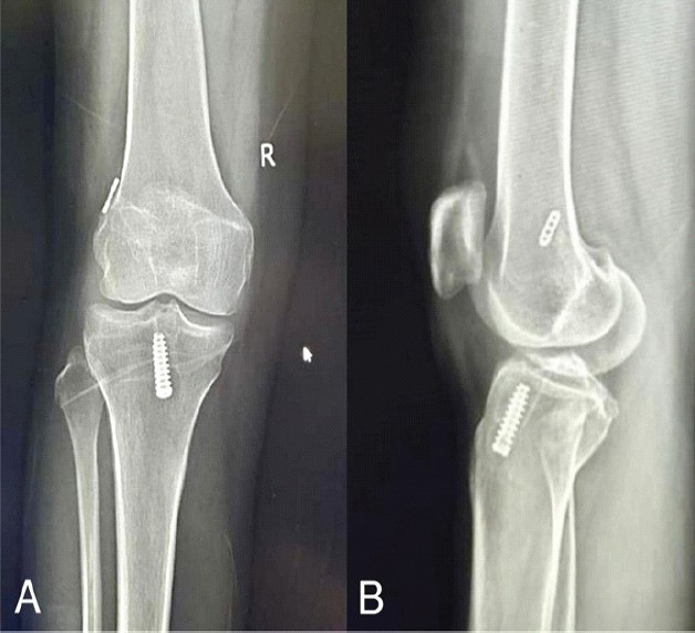
Figure 3. Post-operative X-rays of the knee at 6 months; (a) anteroposterior and (b) lateral X-rays of the knee at 6 months follow-up showing an interference screw within the proximal tibial tunnel and an endobutton on the femoral cortex. Joint space is well-maintained, and there is no evidence of any hardware-related complication.
Discussion
The efficacy of PRP in ACL reconstruction remains controversial, mainly due to the lack of standardization in preparation protocols and application methods. This study emphasizes two key factors: (1) leukocyte reduction (LP-PRP) to minimize intra-articular inflammatory responses and (2) multi-site delivery to optimize biological effects at graft healing regions.
The observed early functional improvement suggests that PRP may primarily influence the early phase of healing, such as cell proliferation and neovascularization. However, previous studies have reported inconsistent results, possibly due to variations in PRP preparation techniques and application methods.
Therefore, standardizing LP-PRP preparation and applying a targeted delivery strategy may help improve consistency and clinical outcomes.
Advantages of the study
Unlike most studies that focus primarily on the efficacy of PRP, the current study highlights a standardized preparation protocol of LP-PRP, its targeted multi-site delivery in arthroscopic ACL reconstruction, and the early favorable functional outcomes of the patients.
Limitations of the study
Limitations include small sample size, absence of a control group, and lack of long-term follow-up. However, the objective of this report is to describe a reproducible and clinically standardized technique providing a practical framework for future clinical application and research rather than establish definitive efficacy.
References
Pawar, S. S., Naskar, A. K., Jogi, H. K., Sirsat, A. D., Bhowmick, L., & Mehta, M. A. (2026). Standardized Preparation and Multi-Site Application of Leukocyte-Poor Platelet-Rich Plasma in Arthroscopic Anterior Cruciate Ligament Reconstruction: A Technical Note with Early Functional Outcomes. Journal of orthopaedic case reports, 16(6), 115–121.
Source: Journal of orthopaedic case reports.