
Scientific Reports, 16 April 2026
Research Background
Hypoxic–Ischemic Encephalopathy (HIE) is a leading cause of long-term neurological impairment in neonates, particularly in preterm infants. Due to the immaturity of the developing brain, preterm infants are highly susceptible to neuroinflammation, oxidative stress, and impaired cerebral perfusion, which can result in severe outcomes such as cerebral palsy and motor dysfunction.
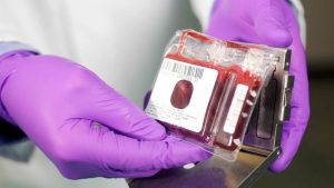
Currently, effective treatment options for HIE in preterm infants remain limited. Therapeutic hypothermia, the standard of care for term infants, is not recommended for preterm populations due to safety concerns and uncertain efficacy. In this context, umbilical cord blood–derived stem cells, particularly the CD34⁺ population, have attracted increasing attention due to their potential to promote angiogenesis and modulate inflammatory responses.
Methods
This preclinical study was conducted using a neonatal mouse model to mimic brain injury in preterm infants. Postnatal day 5 (P5) mouse pups were used, as their stage of brain development is comparable to that of human preterm infants.
Brain injury was induced using a unilateral hypoxia–ischemia model. The left common carotid artery was ligated and transected under isoflurane anesthesia, followed by exposure to hypoxic conditions (8% O₂) for 50 minutes. After recovery, the pups were returned to their dams and monitored.
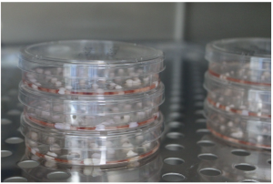
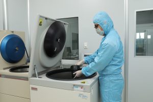
CD34⁺ cells were isolated from human umbilical cord blood and divided into two groups: non-expanded cells and cells expanded ex vivo for 10 days using a fully defined culture system free of serum, cytokines, and albumin. Conventional biological components were replaced with synthetic molecules to maintain the self-renewal and proliferative capacity of hematopoietic stem cells.
At 48 hours post-injury, the animals were randomly assigned to four groups: sham, untreated HIE, HIE treated with CD34⁺ cells, and HIE treated with expanded CD34⁺ cells. Each animal received an intravenous injection of 1×10⁵ cells via the facial vein.
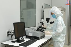
Therapeutic efficacy was comprehensively evaluated through measurements of cerebral blood flow, body weight monitoring, behavioral assessments (negative geotaxis, open-field, and cylinder tests), histological brain analysis, and neuroinflammation assessment using GFAP and Iba1 markers. Phenotypic characterization of CD34⁺ cells before and after expansion was performed by flow cytometry to confirm maintenance of stem cell properties.
Key Results
Both CD34⁺-treated groups demonstrated significant improvement in motor function compared to the untreated HIE group, particularly in the cylinder test, a sensitive indicator of motor cortex injury. In addition, a marked reduction in activated microglia and astrocytes was observed in the injured brain regions, indicating an anti-inflammatory effect.
However, no significant improvements were observed in morphological parameters such as body weight or brain volume. Importantly, administration of CD34⁺ cells, including ex vivo expanded cells, did not increase mortality, supporting the safety profile of this approach.
Notably, the therapeutic efficacy of expanded CD34⁺ cells was comparable to that of non-expanded cells, indicating that the expansion process did not compromise their biological function.
Scientific Interpretation
The findings support the notion that the therapeutic effects of CD34⁺ cells are primarily mediated through paracrine mechanisms, including the secretion of growth factors, anti-inflammatory cytokines, and immunomodulatory molecules. These factors contribute to improving the injured brain microenvironment, reducing inflammation, and facilitating functional recovery.
Ex vivo expansion under defined culture conditions offers a significant advantage by overcoming the limitation of insufficient cell numbers, particularly in preterm infants with limited cord blood volume. Moreover, the elimination of serum and cytokines enhances product consistency and safety, aligning with clinical-grade cell manufacturing standards.
Conclusion and Future Perspectives
Umbilical cord blood–derived CD34⁺ cell therapy, particularly when combined with ex vivo expansion technology, represents a promising strategy for the treatment of brain injury in preterm infants. Intravenous administration at 48 hours post-injury appears to be a feasible and clinically relevant approach.
Further studies are warranted to optimize dosing, timing, and to elucidate the underlying mechanisms of action before translation into clinical applications. These findings provide an important foundation for the development of regenerative therapies targeting neonatal brain injury.
References
Arai N., Tanaka E., Ohnishi S. et al. Intravenous administration of ex vivo expanded human umbilical cord blood-derived CD34⁺cells in a preterm hypoxicischemic encephalopathy mouse model. Sci Rep (2026). https://doi.org/10.1038/ s41598-026-4820
Source: Scientific Reports
Link: https://doi.org/10.1038/ s41598-026-4820